Iron deficiency anemia (IDA)
Iron deficiency anemia (IDA) is one of the most frequently diagnosed pathological conditions of the circulatory system, and indeed the most common type.
Statistical studies have shown that about 2.5 billion patients worldwide have this diagnosis.
In order to stop the progression of the disease and avoid complications, it is necessary to identify the root causes of its occurrence and begin treatment in a timely manner.
What is iron deficiency anemia?
Anemia is characterized by a reduced content of red blood cells - erythrocytes - in the human circulatory system, and, as a result, a drop in hemoglobin.
If the low level of these elements is associated with a lack of iron in the body, then in this case we are talking about iron deficiency anemia (IDA).
As a rule, pathology is not an independent disease. In most cases, iron deficiency anemia occurs following some other negative changes in the human body.
FOR REFERENCE! The average amount in the body for adults is about 4 grams. For men and women at different ages, this indicator may have different meanings. For example, iron deficiency anemia in adults is much more common in the fairer sex. First of all, this is due to regular blood loss that occurs during menstruation. And the strongest concentration of iron is observed in newborn babies, since they have an increased supply of this trace element in the womb.
Iron deficiency has a negative impact on human vitality in general. In addition, the development of this deficiency is fraught with disruptions in the formation of red blood cells, as well as disruption of oxidation and reduction reactions, the mechanism of cell division and the normal course of some other reactions.
Iron is the basis of hemoglobin, which performs the function of supplying oxygen to all tissues and organs in the human body, and also plays an important role in the synthesis of protein and hormones. If iron deficiency is not replenished for a long time, the patient begins to develop anemic syndrome.
Causes of iron deficiency anemia
The reasons for the development of iron deficiency anemia may be a lack of iron entering the body from the outside, or failures in the processes that consume it, because the human body cannot produce this microelement on its own. They could be:
- unbalanced diet: poorly chosen diet, refusal to eat meat (vegetarianism);
- regular significant blood loss . In addition to menstruation in women, chronic blood loss can be associated with the presence of various diseases: decaying tumors and others. This also includes blood donation, which occurs more often than 3-4 times in one year;
- congenital factors that arose during intrauterine development: the presence of iron deficiency anemia in the mother, multiple pregnancy, prematurity;
- malfunctions of the gastrointestinal tract, as a result of which the process of iron absorption in the duodenum is disrupted. This may be due to the presence of various (enteritis, stomach cancer, etc.);
- leading to disturbances in the production of transferrin – a protein that performs transport functions: microelements supplied with food are not distributed throughout the body, which causes iron deficiency. Transferrin synthesis occurs in liver cells;
- taking medications that affect the absorption and processing of iron in excess doses. These may include: antacids, iron-binding drugs. People with a predisposition to iron deficiency anemia should consult a doctor before using these types of medications.
Iron deficiency anemia in children can develop as a result of various pathologies during pregnancy, early transition to artificial feeding, accelerated growth rate (in case of prematurity).
Provoking factors of IDA

The body's increased need for iron is the main provoking factor for the development of iron deficiency anemia. It can be associated with such life processes as:
- pregnancy. During pregnancy, a woman needs almost twice as much iron for normal fetal development as in normal life;
- breast-feeding. As during pregnancy, while feeding a child, the female body uses much more iron than it can receive.
Stages of development of IDA
The pathogenesis of this type of anemia is expressed in two main periods:
- Latent (hidden) period characterized by a decrease in iron reserves in the body, resulting in a decrease in ferritin levels. However, other laboratory parameters may remain within normal limits. The body tries to compensate for the lack of a microelement by more active absorption in the intestines and the production of transport protein. Due to this, IDA has not yet occurred at this stage, although the prerequisites for it are already present.
- Direct iron deficiency anemia occurs at the moment when the level of red blood cells decreases so much that they can no longer sufficiently provide their functions. At this stage, the main symptoms and characteristic features of the disease begin to appear more clearly.
Types of iron deficiency anemia
Classification of the disease according to its causes distinguishes the following types:
- anemia resulting from excessive blood loss;
- iron deficiency anemia, which appeared as a result of malfunctions of red blood cells;
- chronic iron deficiency anemia;
- hemolytic anemia (increases with a high degree of destruction of red blood cells).
Classification according to hemoglobin level divides the disease into types depending on severity:
- mild severity (hemoglobin content more than 90 g/l);
- moderate severity (70-90 g/l);
- high severity (below 70 g/l).
Symptoms of iron deficiency anemia

The degree of IDA increases gradually in the body and at first may hardly make itself felt. The latent period of the disease is characterized by the manifestation of sideropenic syndrome.
Later, a general anemic syndrome begins to appear, the clarity of which is determined by the severity of anemia and the body’s ability to resist. The presence of the following signs in a patient may indicate iron deficiency anemia:
- fatigue and chronic muscle fatigue. With iron deficiency in the body, a person's muscles become weaker. Their daily work requires a lot of energy, which is no longer produced in the required quantity due to a decrease in the level of red blood cells. As a result, a person gets tired much faster even with small everyday loads. Iron deficiency anemia in children can manifest itself in the child's desire for less active play, lethargic behavior and drowsiness;
- the appearance of shortness of breath. With IDA, it is difficult to supply the heart with oxygen due to deterioration of blood circulation. For this reason, the patient may experience shortness of breath;
- deterioration of the condition of the skin, nails and hair. Iron deficiency becomes noticeable externally (see photo above), when the skin becomes dry and cracked, and pallor appears. Nails weaken, break and become covered with specific transverse cracks. In some cases, the nail plate may bend in the opposite direction. The hairline is thinning. Hair changes its structure, gray hair appears prematurely;
- damage to the mucous membranes. One of the early symptoms of IDA is damage to the mucous membranes, because these tissues most acutely feel the lack of iron due to disruption of various cellular processes:
- Damage to the mucous membranes is most clearly noticeable in changes in the appearance of the tongue. It becomes smooth, covered with cracks and areas of redness. Pain and burning sensations are added. In some cases there are sharp;
- dryness and areas of atrophy appear in the oral cavity. There is discomfort when eating food and pain when swallowing. Cracks form on the lips;
- atrophy of the intestinal mucosa caused by iron deficiency anemia is accompanied by the appearance of pain syndromes in the abdomen, constipation, and diarrhea. The ability of the gastrointestinal tract to absorb nutrients deteriorates;
- damage to the mucous membranes of the genitourinary system is characterized by pain during urination and (usually in childhood). The risk of contracting various infections increases;
- susceptibility to various infections. A lack of iron in the body also affects the work of leukocytes - blood cells responsible for freeing the body from pathogens of various infections. As a result, the patient experiences a general weakening of the immune system and increases susceptibility to bacterial and viral infections;
- difficulties with intellectual activity. Insufficient supply of brain cells with iron leads to memory impairment, absent-mindedness and weakening of intelligence in general.
Diagnosis of IDA
Iron deficiency anemia in children and adults can be diagnosed by any specialist, however, detailed diagnosis aimed at identifying the causes and treatment should be carried out by a hematologist. Patient examinations include:
- Visual examination of the patient is the first stage of diagnosing IDA. The specialist needs, from the patient’s words, to determine the general picture of the development of the pathology and conduct an examination that will help draw conclusions about the extent of the disease and identify complications, if any;
- from a finger or from a vein - a generalized picture of the patient’s health, with the help of which the doctor can unambiguously determine the presence or absence of IDA in the patient. This analysis is carried out in the laboratory using special equipment - a hematology analyzer. The diagnosis of iron deficiency anemia is established in a patient if:
- a decrease in the number of red blood cells (in men - less than 4.0 x 10¹²/l, in women - less than 3.5 x 10¹²/l), when the number of platelets and leukocytes is normal or increased;
- the predominance of red blood cells in the patient’s blood, the size of which is less than normal (a deviation is considered to be a size of less than 70 µm3);
- color index (CI) is less than 0.8;
- a biochemical blood test allows a more detailed study of the patient’s condition, taking into account indicators related to the area under study. The following abnormalities indicate the presence of iron deficiency anemia:
- serum iron (SI): in men – less than 17.9 µmol/l, in women – less than 14.3 µmol/l;
- total serum iron binding capacity (TIBC): significantly exceeds the level of 77 µmol/l;
- ferritin (a complex protein complex that acts as the main intracellular iron depot in humans) is below normal: in men - below 15 ng/ml, in women - less than 12 ng/ml;
- (less than 120 g/l);
Iron deficiency anemia in children is characterized by the following blood test results:
- serum iron (SI) below 14 µmol/l;
- total iron binding capacity of serum (TIBC) more than 63 µmol/l;
- ferritin in the blood is below 12 ng/ml;
- hemoglobin level (less than 110 g/l).
- Bone marrow puncture is a diagnostic method based on the collection of bone marrow samples by taking it with a special instrument from the sternum. With IDA disease, an increase in the erythroid lineage of hematopoiesis is observed;
- X-rays are carried out to determine intestinal pathologies that can cause chronic bleeding, thereby causing the development of anemia;
- endoscopic examinations of human mucous membranes are also carried out to identify various pathologies of the abdominal organs. It can be:
- fibroesophagogastroduodenoscopy (FEGDS);
- sigmoidoscopy;
- colonoscopy;
- laparoscopy and others.
Treatment of iron deficiency anemia
According to doctors, when treating iron deficiency anemia in adults and children, one cannot limit oneself only to medications. It is best and easiest to compensate for the deficiency of an important microelement with the help of healthy food and a properly selected diet.
The daily requirement of iron that the diet should contain is at least 20 mg. It should be noted that treatment of this disease will be ineffective if measures are not taken to eliminate the primary pathology that caused iron deficiency.
To prevent the disease, each person should undergo a laboratory analysis of blood counts every year, eat a comprehensive diet and, if necessary, promptly eliminate possible causes of significant blood loss.
People with a predisposition to iron deficiency should consult a doctor for a course of medications with high iron content.
Nutrition and supplements

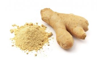
A balanced diet plays a major role in the prevention and treatment of IDA. When planning a diet, it should be taken into account that iron is better absorbed if it is taken in conjunction with vitamin C.
Moreover, this microelement is best absorbed by the intestines if it is contained in products of animal origin (up to 3 times more compared to plant products).
- white beans (72 mg);
- nuts of all types (51 mg);
- buckwheat (31 mg);
- pork liver (28 mg);
- molasses (20 mg);
- brewer's yeast (18 mg);
- seaweed and seaweed (16 mg);
- pumpkin seeds (15 mg);
- lentils (12 mg);
- blueberries (9 mg);
- beef liver (9 mg);
- heart (6 mg);
- beef tongue (5 mg);
- dried apricots (4 mg).
- ascorbic acid;
- succinic acid;
- fructose;
- nicotinamide
FOR REFERENCE! Seafood is also rich in iron, but it is not recommended to include it in the diet if you are deficient in this microelement. The fact is that, among other things, they contain a large amount of phosphates, which complicate the process of absorption of iron in the body.
Despite the fact that iron deficiency anemia rarely develops in infants (except when the mother has this disease), it should be noted that in this case the disease is especially dangerous.
A lack of iron in children can result in serious impairments in physical development, and therefore requires prompt replenishment.
Treatment of iron deficiency anemia at this age is carried out through a strict diet and careful monitoring of the baby's daily intake, as well as a review of the baby's complementary feeding, if it is already available.
Drug treatment (medicines)

Proper nutrition is a necessary step in the treatment and prevention of IDA, but it cannot compensate for the lack of the necessary microelement in the body on its own, and therefore doctors recommend that patients take medications.
Most often, medications are prescribed in the form of tablets; less often, in cases of intestinal dysfunction, parenteral administration is prescribed.
Medicines for iron deficiency anemia should be taken over a long course (over several weeks or months).
All of them are designed to normalize the main indicators in a blood test and eliminate the symptoms of the disease. The most commonly used of them:
- Hemophere prolongatum;
- Sorbifer Durules;
- Ferroceron;
- Ferroplex;
- Tardiferon.
Before using medications, you should consult your doctor; improper use can lead to an excess of iron, which is also fraught with negative consequences and complications.
Red blood cell transfusion
In complex cases of anemia, it may be necessary to undergo a red blood cell transfusion. This procedure may be necessary if there is a serious threat to the patient’s life and should be carried out in the shortest possible period of time. Indications for prescribing red blood cell transfusion may be:
- significant blood loss;
- a sharp decrease in hemoglobin levels;
- preparation for surgery or early childbirth.
For the successful implementation of this procedure and the absence of complications, it is very important that the donor’s blood ideally matches the patient in all laboratory parameters.
Prognosis and complications
The degree of complexity of such a disease as iron deficiency anemia is quite low today.
With timely detection of symptoms and high-quality diagnosis, this disease can be completely eliminated without any consequences.
In some cases, complications may develop during the treatment of IDA. The reasons for this may be the following factors:
- illiterate implementation of diagnostic procedures and, as a result, establishment of a false diagnosis;
- failure to identify the first cause;
- untimely adoption of treatment measures;
- incorrect dosage of prescribed medications;
- non-compliance with regularity of treatment.
Possible complications of this disease are:
- in children – growth retardation and intellectual development. Children's iron deficiency is very dangerous, since in advanced cases of the disease, disruptions in the child's body can become irreversible;
- anemic coma, which develops against the background of poor-quality oxygen circulation in the body, in particular, due to insufficient oxygen supply to the brain. Characteristic signs of this complication are fainting, weakened and diminished reflexes. Failure to provide timely qualified medical assistance creates a strong threat to the patient’s life;
- appearance is a common occurrence with a long-term lack of iron in the body;
- Infectious diseases with anemia can cause the development.
These types of complications pose the greatest threat to pediatric and elderly patients.
Related videos
Interesting
For citation: Dvoretsky L.I. IRON DEFICIENCY ANEMIA. breast cancer. 1997;19:2.
AND
Iron deficiency anemia (IDA) is defined as a clinical and hematological syndrome, which is based on a violation of hemoglobin synthesis due to iron deficiency, which develops in various pathological (physiological) processes. Along with the developed symptom complex of IDA, the so-called hidden iron deficiency is distinguished, characterized by a decrease in the iron content in depots (reserves) and serum while maintaining normal hemoglobin levels. Latent iron deficiency is a prestage of IDA, which develops with further progression and lack of compensation.
IDA is the most common anemic syndrome and accounts for approximately 80% of all anemias. According to WHO, the number of people with iron deficiency reaches 200 million worldwide. The population groups most vulnerable to the development of IDA include young children, pregnant women, and women of childbearing age. In developed countries of Europe and Russia, about 10% of women of childbearing age suffer from IDA, and 20% of women have hidden iron deficiency. The frequency of iron deficiency conditions in the form of hidden iron deficiency in some regions of Russia (North, Eastern Siberia, Northern Caucasus) reaches 50-60%. The prevalence of IDA in children in Russia and developed European countries is about 50%.
Clinical picture
Clinical manifestations of IDA are caused, on the one hand, by the presence of anemic syndrome, and on the other, by iron deficiency (hyposiderosis).
Anemic syndrome is manifested by well-known and nonspecific symptoms for anemia of any origin (dizziness, tinnitus, spots before the eyes, shortness of breath, palpitations, etc.). In most cases, the decrease in hemoglobin level occurs gradually (unlike acute blood loss), while various organs adapt to anemia, and therefore the complaints of patients do not always correspond to the hemoglobin level. Many patients, especially women, get used to their illness, attributing it to overwork, mental and physical overload. Often, patients turn to or see a doctor for the first time due to unexpected and worrying situations such as fainting, associated falls, as well as long-term asthenia and decreased performance after viral and other respiratory infections. With a decrease in hemoglobin levels in patients with coronary heart disease (CHD), angina attacks may become more frequent, the need for nitroglycerin increases, and exercise tolerance decreases. In some cases, angina complaints are leading in the clinical picture, and therefore patients are hospitalized for unstable angina or suspected myocardial infarction. In the presence of severe anemia, signs of heart failure may appear, characterized by an increase in minute blood volume (anemic heart), and in cases of pre-existing heart failure, the latter may worsen against the background of the development of anemia and become refractory to treatment. In patients with discirculatory encephalopathies, especially in old age, against the background of the development of IDA and tissue hypoxia of the brain, decompensation of the existing vascular lesion of the brain occurs.
hyposiderosis syndrome. Clinical manifestations of hyposiderosis are associated with tissue deficiency of iron, which is necessary for the functioning of organs and tissues. The main symptoms of hyposiderosis are observed on the part of epithelial tissues (skin and its appendages, mucous membranes) as a result of a decrease in the activity of some iron-containing tissue enzymes, in particular cytochromes. Dry skin and impaired integrity of the epidermis are noted. Ulcerations and cracks with an inflammatory shaft (angular stomatitis) appear in the corners of the mouth. Typical clinical manifestations of hyposiderosis are brittleness and layering of the nails, their transverse striations. The nails become flat, sometimes taking on a concave spoon-shaped shape (koilonychia).
Some patients report a burning sensation on the tongue. A perversion of taste is possible in the form of an indomitable desire to eat chalk, toothpaste, ashes, and the like, as well as an addiction to certain odors (acetone, gasoline). The morphological substrate of manifestations of hyposiderosis on the part of the oral mucosa is atrophy, hyperkeratosis, vacuolization of the epithelium with a sharp decrease in the content of respiratory enzymes (cytochrome oxidase and succinate dehydrogenase) in epithelial cells. One of the signs of hyposiderosis is difficulty swallowing dry and solid foods (sideropenic dysphagia), which forces the doctor to suspect a tumor lesion of the esophagus. In girls, and less often in adult women, dysuric disorders are possible, sometimes urinary incontinence when coughing or laughing, which gives a urological focus to the examination of such patients. Children may experience symptoms of nocturnal enuresis. Dystrophic changes in the cells of the gastric mucosa, mainly its body, occur, with the development in some cases of secretory insufficiency and the appearance of corresponding clinical symptoms (feeling of heaviness, pain), which are not as pronounced as with gastritis of other origins.
Symptoms associated with iron deficiency include muscle weakness, which is observed in most patients with IDA and is associated not only with anemia, but also with a deficiency of iron-containing enzymes.
When examining patients, attention is drawn to the pallor of the skin, which often has an alabaster or greenish tint. Hence the old name for this type of anemia - chlorosis (greenness). Often in patients with IDA there is a distinct “blue” discoloration of the sclera (blue sclera symptom). It is believed that the sensitivity and specificity of this sign are 89 and 64%, respectively. This phenomenon is explained by the fact that with iron deficiency, dystrophic changes in the cornea of the eye occur, through which the choroid plexuses are visible, creating a “blue” appearance. This sign, which attracts attention when examining patients with anemia, allows the doctor to suspect the iron deficiency nature of anemia and determine the direction of the diagnostic search.
Laboratory signs of IDA. The main laboratory sign that allows one to suspect the iron deficiency nature of anemia is a low color index, which reflects the hemoglobin content in the erythrocyte and is a calculated value. Since in IDA, hemoglobin synthesis is impaired due to a lack of “building” material, and the production of red blood cells in the bone marrow is reduced slightly, the calculated color index is always below 0.85, often 0.7 or less (all IDA are hypochromic!).
When using modern analyzers in laboratory practice, it is possible to directly determine the average hemoglobin content in one erythrocyte (MCH; normal 27-35 pg) and the average hemoglobin content in erythrocytes (MCHC; normal 31-36 g per 100 ml of blood).
Morphologically, in hypochromic anemia, hypochromic erythrocytes are detected, which predominate in the peripheral blood smear and are characterized by the presence of wide clearing in the center of the erythrocyte.
The red blood cell resembles a donut or a ring (anulocyte). In addition, in the blood smear of patients with IDA, microcytes are often found, in which the hemoglobin content is less than in normal-sized red blood cells.
In a peripheral blood smear, along with microcytosis, anisocytosis and poikilocytosis are noted, i.e., red blood cells of unequal size and different shapes are found, the number of siderocytes (red blood cells with iron granules detected by special staining) is sharply reduced compared to the norm, up to their complete absence. The content of reticulocytes in the blood, as a rule, is within normal limits, with the exception of cases of severe blood loss due to corresponding pathology (excessive nasal and uterine bleeding) or during treatment with iron supplements. The number of leukocytes and platelets is usually unchanged. Some patients may experience thrombocytosis, which disappears after correction of IDA.
Morphological examination of the bone marrow for diagnosing IDA is not very informative and can only be of value with special staining for iron and counting sideroblasts (erythroid cells of the bone marrow with iron granules), the number of which is significantly reduced in patients with IDA.
The iron content in blood serum taken before the start of iron therapy is reduced, often significantly. Normal serum iron levels in men and women are 13-30 and 12-25 µmol/l, respectively. Along with determining the concentration of iron in the serum, the assessment of the total iron binding capacity of the serum (TIBC), which reflects the degree of “starvation” of the serum and saturation of transferrin with iron, is of diagnostic importance. The method consists in adding a known excess of iron to the subject’s serum, part of which binds to protein, and the other, unbound part is removed by absorption on an ion exchange resin. After this, the content of iron bound to the protein is determined and the amount of iron that can bind 1 liter of whey is calculated. This indicator reflects the total blood pressure (normally 30-85 µmol/l). The difference between TBI and serum iron values reflects the latent iron-binding capacity, and the ratio of serum iron to TBI, expressed as a percentage, reflects the percentage of transferrin saturation with iron (normal 16-50%).
In patients with IDA, there is an increase in CVB, a significant increase in latent iron-binding capacity and a decrease in the percentage of transferrin saturation.
Since iron reserves are depleted in IDA, there is a decrease in the serum content of ferritin, an iron-containing protein, the level of which, along with the concentration of hemosiderin, reflects the amount of iron reserves in the depot. A decrease in serum ferritin levels is the most sensitive and specific laboratory sign of iron deficiency and confirms the iron deficiency nature of the anemic syndrome. Normal ferritin levels average 15-150 µg/l ( menstruating women have lower rates than men). An assessment of iron stores can be carried out by determining the iron content in the urine after the administration of certain complexes that bind iron and excrete it in the urine. Desferal (desferoxamine) is used for this purpose. After intravenous administration of 500 mg of desferal, 0.8 to 1.2 mg of iron is normally excreted, while in patients with IDA or in the presence of hidden iron deficiency, the amount of iron excreted in the urine is reduced to 0.2 mg or less. At the same time, with excess iron content in the depot, in some anemias in which iron is not used by erythroid cells, the amount of iron excreted in the urine after the administration of desferal exceeds the norm.
Another way to assess iron stores is to stain blood and bone marrow smears for iron and count the number of siderocytes and sideroblasts. The number of these cells in IDA is significantly reduced.
Diagnosis of IDA
The diagnostic search for suspected IDA can be conventionally presented in the form of several successive stages.
1. Diagnosis of hypochromic anemia represents the most important stage, since it is the hypochromic nature of anemia that is the key sign that allows one to suspect, first of all, IDA (all IDA are hypochromic!) and determine the further direction of the diagnostic search. In this regard, when interpreting the results of a blood test, a clinician must pay attention not only to the color indicator (it may be calculated incorrectly if the laboratory assistant incorrectly counts the number of red blood cells), but also to the morphological picture of red blood cells, which is reflected in the analysis by the laboratory doctor examining smear (for example, hypochromia, microcytosis, etc.).
2. Differential diagnosis of hypochromic anemia. The presence of hypochromic anemia makes it very likely to assume the presence of iron deficiency anemia, which forms the main group among hypochromic anemias, but does not exclude hypochromic anemias of other origins (not all hypochromic anemias are iron deficiency!). In this regard, at this stage of the diagnostic search, it is necessary to carry out a differential diagnosis between IDA and the so-called sideroachrestic (achresia - non-use) anemia. With sideroachrestic anemia (a group concept), also referred to as iron-saturated anemia, the iron content in the body is within normal limits or there is even an excess.
However, for various reasons, iron is not used to build heme in the hemoglobin molecule, which ultimately leads to the formation of hypochromic red blood cells with a low hemoglobin content. Unused iron enters reserves and is deposited in organs and tissues (liver, pancreas, skin, macrophage system, etc.), leading to the development of hemosiderosis.
Correctly recognizing IDA and distinguishing it from sideroachrestic anemia is extremely important, since an erroneous diagnosis of IDA in patients with iron-saturated anemia may lead to unjustified prescription of iron supplements to these patients, which in this situation will lead to an even greater “overload” of organs and tissues with iron, while there will be no therapeutic effect.
The main types of hypochromic anemia with which the differential diagnosis of IDA should be made:
- anemia associated with impaired heme synthesis, resulting from inhibition of the activity of certain enzymes (heme synthetase), which ensure the inclusion of iron in the heme molecule. This enzyme defect may be of a hereditary nature (hereditary sideroachrestic anemia) or occur as a result of exposure to certain medications (isoniazid, PAS, etc.), alcohol intoxication, contact with lead, etc.
Hypochromic anemia may be one of the manifestations of chronic lead intoxication, in which the synthesis of porphyrins, an integral part of the heme molecule, is impaired;
- thalassemia, which belongs to the group of hereditary hemolytic anemias associated with impaired synthesis of globin - the protein part of hemoglobin. The disease has several variants and is characterized by signs of hemolysis (reticulocytosis, increased levels of indirect bilirubin, enlarged spleen), high iron content in the serum and in the depot, and hypochromic anemia. In fact, with thalassemia we are also talking about sideroachresia, i.e. about the lack of use of iron, but not as a result of defects in the enzymes involved in heme synthesis, but due to a disruption in the process of constructing the hemoglobin molecule as a whole due to the pathology of its globin part;
- anemia associated with chronic diseases. This term is usually used to designate a group of anemias that occur in patients against the background of various diseases, most often of an inflammatory nature (infectious and non-infectious).
An example is anemia in suppurative diseases of various locations (lungs, abdominal cavity, osteomyelitis), sepsis, tuberculosis, infective endocarditis, rheumatoid polyarthritis, malignant tumors in the absence of blood loss. With all the diversity of pathogenetic mechanisms of anemia in these situations, one of the main ones is considered to be the redistribution of iron into the cells of the macrophage system, which is activated when inflammatory and tumor processes. Since true iron deficiency is not observed in these anemias, it is more justified to talk not about IDA, but about iron redistribution anemia. The latter are, as a rule, moderately hypochromic in nature, the iron content in the serum may be slightly reduced, the life-sustaining blood pressure is usually within the normal range or moderately reduced, which distinguishes this variant of anemia from iron deficiency anemia. An increase in the level of ferritin in the blood is characteristic. Understanding and correct interpretation of the pathogenetic mechanisms of anemia development in the above diseases allows the doctor to refrain from prescribing iron supplements to these patients, which are usually ineffective.
Thus, we can talk about the presence of IDA in cases of hypochromic anemia, accompanied by a decrease in serum iron content, an increase in PVSS, and a decrease in ferritin concentration. To avoid errors when interpreting the results of determining the iron content in serum, the doctor must take into account a number of factors that influence the obtained indicators:
if the study is carried out after taking iron supplements (even for a short period of time), then the obtained indicators do not reflect the true iron content in the serum. In this regard, the study should be carried out before starting treatment with iron preparations.
If the latter were prescribed, then the study can be carried out no earlier than 7 days after their cancellation;
- red blood cell transfusions, often carried out before the nature of anemia is clarified (marked decrease in hemoglobin levels, signs of heart failure, etc.), also distort the assessment of the true iron content in the serum;
- To test serum for iron content, special test tubes should be used, washed twice with distilled water, since the use of tap water containing small amounts of iron for washing affects the results of the study. Drying cabinets should not be used to dry test tubes, since a small amount of iron gets into the dishes from their walls when heated;
- Currently, for the study of iron, it is customary to use bathophenanthraline as a reagent, which forms a colored complex with iron ions with a stable color and a high molar extinction coefficient; the accuracy of the method is quite high;
- Blood for analysis should be taken in the morning, since there are daily fluctuations in the concentration of iron in the serum (iron levels are higher in the morning). In addition, it must be borne in mind that serum iron levels are influenced by the phase of the menstrual cycle (immediately before and during menstruation, serum iron levels are higher), pregnancy (increased iron levels in the first weeks of pregnancy), and the use of oral contraceptives (increased ), acute hepatitis and liver cirrhosis (increased). Random variations in the studied parameters may be observed.
3. Identifying the cause of IDA. After confirming the iron deficiency nature of anemia, i.e., verification of IDA syndrome, an equally important task is to establish the cause of this anemic syndrome. Recognizing the cause of the development of IDA in each specific case is the final stage of the diagnostic search. Focus on nosological diagnosis is very important, since in most cases, when treating anemia, it is possible to influence the underlying pathological process.
Causes of IDA
The main reasons for the development of IDA are chronic blood loss, impaired absorption in the intestine, increased need for iron, impaired iron transport, and nutritional deficiency. Each of these causes is usually characteristic of a certain group of patients with IDA and occurs in appropriate clinical situations. Thus, an increased need for iron underlies IDA in pregnant and lactating mothers. In menstruating women, the main cause of IDA is menorrhagia, and in children it is nutritional deficiency.
Chronic blood loss occupies the main place among the causes of IDA. These blood losses are usually characterized by a small volume of blood lost, short duration, often occur unnoticed by patients and are not always adequately assessed as a cause of IDA by doctors of various specialties. Doctors often forget about the various mechanisms of anemia development during acute and chronic blood loss or underestimate these mechanisms. If during acute blood loss anemia develops as a result of a decrease in the mass of red blood cells and depends both on the degree of blood loss and on the compensatory activation of erythropoiesis, then chronic blood loss (even small in volume, but relatively long-term) leads over time to depletion of iron reserves with the subsequent development of IDA. If we assume that 1 ml of blood contains 0.5 mg of iron, then the daily loss of 2-3 teaspoons of blood (10 ml, i.e. 5 mg of iron) if a patient has, for example, mole-bearing hemorrhoids, exceeds the daily intake of iron, which depletes its reserves and is a risk factor for IDA.
The main sources of chronic blood loss that can lead to the development of IDA are as follows.
1.
Gastrointestinal tract (GIT). Blood loss from the gastrointestinal tract is the most common cause of IDA in men and non-menstruating women; they can occur with various diseases throughout the gastrointestinal tract:
- bleeding from the gums;
- erosive esophagitis (often due to reflux with cardiac insufficiency);
- varicose veins of the esophagus and cardia of the stomach (with cirrhosis of the liver and other forms of portal hypertension);
- acute and chronic gastric erosions (often medicinal in nature);
- peptic ulcer of the stomach and duodenum;
- stomach tumors (usually malignant);
- tumors of the small intestine (rare);
- diverticulosis of the small intestine (Meckel's diverticulum);
- terminal ileitis (Crohn's disease);
- diverticular bowel disease (often with diverticulitis);
- nonspecific ulcerative colitis;
- bleeding hemorrhoids.
Recognizing the source of chronic blood loss requires the doctor to thoroughly examine the gastrointestinal tract (in some cases, repeatedly) using modern methods (x-ray, ultrasound, endoscopic, radioisotope, etc.).
Sometimes the source of chronic blood loss from the gastrointestinal tract can be Meckel's diverticulum, which is a congenital anomaly (defect in the development of the bile duct) and is localized in the small intestine, usually at a distance of 10-20 cm from the cecum. The mucous membrane of the diverticulum sometimes resembles the gastric mucosa and produces hydrochloric acid and pepsin, which causes ulcers to form and bleeding occurs, leading to the development of IDA.
Symptoms from the abdominal organs are nonspecific and often absent altogether. The source of bleeding can only be identified during laparotomy.
2. Uterine blood loss are the main cause of IDA in women of childbearing age and can be observed in the following conditions:
- menorrhagia of various origins (platelet dysfunction, etc.);
- dysfunctional uterine bleeding;
- uterine fibroids;
- endometriosis;
- malignant tumors of the uterus;
- presence of intrauterine contraceptives;
- retained placenta.
A large group of women suffering from menorrhagia, in whom a gynecologist does not detect any pathology during examination and the cause of menorrhagia remains unclear, deserves special attention.
Having received from the gynecologist the conclusion “there is no data indicating the presence of gynecological pathology,” confirming the absence of a connection between anemia and existing menstrual blood loss, the general practitioner begins a new cycle of examination of the patient in an attempt to establish the true nature of the anemic syndrome. Meanwhile, a simple calculation of the approximate amount of iron lost in menstrual blood allows us to assess the true clinical significance of menorrhagia in the development of IDA in the absence of compensation for these losses. Thus, the average menstrual blood loss is about 50 ml (25 mg of iron), which determines additional iron losses (about 1 mg per day) compared to men. At the same time, it is known that in women suffering from menorrhagia of various origins, the amount of blood lost during one menstruation reaches 200 ml (100 mg of iron) or more and, therefore, the daily loss of iron is about 4 mg. In such situations, the loss of iron in 1 day already exceeds its intake by 1 mg, in 1 month - by 30 mg, and in 1 year, iron deficiency reaches 360 mg. It is not difficult to understand that in the context of ongoing menorrhagia, in the absence of compensation for iron losses and as its reserves are depleted, women develop iron deficiency and subsequently IDA. The timing of the development of IDA depends on the severity of menorrhagia, the amount of initial iron reserves, and the presence of other risk factors for the development of IDA. Taking this into account, the internist, when identifying the causes of anemia in women of childbearing age, must obtain information about the duration of menstruation (number of days), its intensity (presence of clots, number of pads changed, etc.), duration of the cycle (number of days), duration of presence menorrhagia (months, years).
These issues should be discussed with your gynecologist in an attempt to find optimal ways to manage such patients.
3. Blood loss into closed cavities. Most often we are talking about endometriosis - ectopic growth of the endometrium, most often in the muscular and submucosal layer of the uterus, less often - extragenitally (lungs, gastrointestinal tract, etc.). The cyclical changes that occur in the foci of endometrial tissue lead to bleeding into closed cavities, for example, between the muscular and submucosal layers or inside the muscular layer of the uterus. In this case, the iron shed in the blood is not reused for erythropoiesis and iron deficiency is formed. In some cases, ectopic foci of the endometrium communicate with the uterine cavity, and therefore menorrhagia is observed.
Blood loss into closed cavities is also observed in isolated pulmonary siderosis and so-called glomic tumors.
Isolated pulmonary siderosis is based on damage to the basement membrane of the alveoli. At the same time, red blood cells enter the cavity of the alveoli, absorbed by alveolar macrophages, which contain hemosiderin and are detected in large quantities in the alveoli, alveolar ducts, and interstitial tissue. The anemia that occurs in these patients is of a true iron deficiency nature, since the iron absorbed by macrophages is not utilized for erythropoiesis. The disease can be suspected in young patients with hypochromic anemia, combined with hemoptysis (optional sign), sometimes fever, and X-ray signs of diffuse lung damage (small or large focal shadows against the background of mesh compaction of the lung tissue). Detection of hemosiderin in sputum or bronchoalveolar fluid can provide significant assistance in diagnosis when excluding secondary pulmonary hemosiderosis (mitral stenosis, congenital heart disease). The combination of pulmonary siderosis with kidney damage resembling the picture of glomerulonephritis is called Goodpasture's syndrome.
Glomic tumors arise in the closing arteries found in some arteriovenous anastomoses, for example in the lungs, pleura, intestines, and stomach. These tumors, especially when ulcerated, can lead to blood loss and the development of IDA.
4. Nosebleeds are the cause of the development of IDA mainly in patients with hemorrhagic diathesis (hereditary hemorrhagic telangiectasia, thrombocytopenic purpura).
5. Hematuria how the causes of IDA can occur in chronic hematuric nephritis, IgA nephropathy, urolithiasis, intravascular permanent hemolysis (Marchiafava disease). It should be borne in mind that hematuria does not always clinically manifest itself as gross hematuria and is detected only by examining urine sediment, in particular by staining for hemosiderin if hemoglobinuria is suspected.
6
. Towards the development of IDA can also lead to the so-called iatrogenic blood loss, including frequent blood sampling for research, bloodletting in patients with erythremia and erythrocytosis, blood loss during the hemodialysis procedure in patients with chronic renal failure.
It is possible for donors to develop IDA, especially in the presence of other risk factors (menorrhagia, chronic infections, etc.). In a certain category of patients, mainly in psychiatric practice, IDA can develop due to artificially induced bleeding, most often from the urogenital tract.
7
. Iron malabsorption. Since iron absorption occurs in the duodenum and proximal small intestine, all pathological processes in these parts of the intestine can lead to the development of iron deficiency. The main ones among them are:
- enteritis of various etiologies with the development of malabsorption syndrome;
- resections of the small intestine for various diseases (obstruction, tumors, etc.), leading to a decrease in the area of iron absorption;
- Gastric resection using the Billroth II method (end to side), when part of the duodenum is turned off.
Identifying the above conditions, as a rule, does not present any particular difficulties for the doctor; they can be recognized based on the clinical picture or anamnestic information.
8. Increased need or increased consumption of iron. This cause of IDA usually occurs during pregnancy, lactation, and during periods of intensive growth in girls and adolescents (less often).
In pregnant women, the most common cause of anemia is iron deficiency, especially with repeated and frequent pregnancies and multiple pregnancies. Often, IDA develops in women who give birth less than 3 years apart, since during this period the additional iron costs of the previous pregnancy are not compensated for. Sometimes the hidden iron deficiency that women have before pregnancy manifests itself during pregnancy into a full-blown picture of IDA. The risk of developing IDA in pregnant women is higher in the presence of other risk factors (nutritional deficiency, chronic blood loss, etc.). Along with iron deficiency and, more rarely, folic acid deficiency, the cause of a decrease in hemoglobin levels in pregnant women may be hemodilution due to fluid retention (increased secretion of LDH, aldosterone, etc.). In this case, there is usually no hypochromia of erythrocytes, the iron content in the serum is within normal limits or moderately reduced. Long and frequent lactation can also lead to the development of IDA, especially in the presence of other risk factors.
In clinical practice, there are cases of IDA in girls, less often in adolescents, who do not have chronic blood loss, signs of impaired intestinal absorption and an infectious-inflammatory process. At the same time, these patients experience asthenic manifestations, some developmental delay, and frequent illnesses in childhood. In the past, these anemia variants were referred to as early chlorosis. The studies made it possible to establish that the mothers of these patients suffered from IDA during pregnancy, the treatment of which was inadequate or not carried out at all. In this regard, the fetus received an insufficient amount of iron and the children born had a hidden deficiency, which did not manifest itself until the body experiences an increased need for iron (intensive growth of organs and tissues, the appearance of menstrual blood loss in girls, etc.).
An increased need for iron or its relative deficiency can be observed in patients with B12-deficiency anemia during treatment with vitamin B12, when, when intensive normoblastic hematopoiesis occurs, an amount of iron is required that exceeds the available reserves.
Impaired transport of iron from the blood, leading to the development of IDA, can occur when the level of transferrin, a protein that binds to iron to transfer it into the hemoglobin molecule, decreases in the blood. Similar situations can arise with hypoproteinemia of various origins (nephrotic syndrome with severe proteinuria, impaired protein-synthetic function of the liver, malabsorption syndrome, nutritional deficiency), in which the level of not only albumin, but also globulins, which include transferrin, decreases.
A pronounced decrease in transferrin concentration may be genetic in nature.
9. Nutritional deficiency contributes to the occurrence of IDA due to insufficient intake of iron from food, as well as low protein intake. Such disorders may be important in patients with a low socio-economic standard of living, vegetarians, and in patients with mental anorexia.
Treatment of IDA
When establishing the cause of IDA, the main therapeutic measures should be aimed at eliminating the identified cause (treatment of enteritis, surgical treatment of uterine fibroids, intestinal tumors, etc.). In some cases, the disease underlying IDA does not respond well to radical treatment (hemorrhagic telangiectasia, menorrhagia), and therefore it is necessary to limit oneself to pathogenetic therapy. The basis of pathogenetic therapy for IDA is the use of iron medications orally or parenterally. In the vast majority of cases, in the absence of special indications, iron supplements should be prescribed orally.
To restore hemoglobin levels in patients with IDA, it is necessary that the daily dose of ferrous iron (only it is absorbed) is 100-300 mg, taking into account depleted iron reserves (about 1.5 g). Individual fluctuations are determined by the rate of erythropoiesis, the degree of depletion of iron stores and a number of other factors. In this regard, when choosing an iron preparation and its daily dosage, you should focus not only on the total iron content in it, but mainly on the amount of ferrous iron contained in this preparation. The table presents the main medicinal preparations of iron, the content of other components in them, the amount of total and divalent iron, and the daily dosage of the drug are indicated.
| Basic oral iron preparations | ||||
| A drug | Components | Amount of Fe, mg | Dosage form | Daily dose, g |
| Conferon | succinic acid | Pills | 3-4 | |
| Heferol | Fumaric acid | Capsules | 1-2 | |
| Hemoferprolongatum | Ferrous sulfate | Dragee | 1-2 | |
| Ferrogradumet | Plastic matrix - gradation | Pills | 1-2 | |
| Aktiferrin | D, L-serine |
113,8 |
Capsules Syrup |
1-2
1 teaspoon per 12 kg body weight |
| Ferroplex | Ascorbic acid | Dragee | 8-10 | |
| Sorbifer-durules | “ “ | Pills | 1-2 | |
| Tardiferron | The same + mucoprotease | “ | 1-2 | |
| Fenyuls | Ascorbic acid, nicotinamide, B vitamins | Capsules | ||
| Ferol | Folic acid | 3-4 | ||
| Irovit | The same + ascorbic acid, cyanocobalamin, L-lysine | Capsules | 1-2 | |
| Irradian | Ascorbic acid, folic acid, cyanocobalamin, L-cysteine, D-fructose, yeast | “ | 1-2 | |
It is preferable to prescribe drugs with a higher content of ferrous iron due to the ease of administration for patients (1-2 times a day). The components of many medicinal forms of iron (ascorbic and succinic acids, fructose, cysteine, etc.) enhance the absorption of iron. Iron supplements should be taken with meals for better tolerance. It must be taken into account that under the influence of certain substances contained in food (phosphoric acid, phytin, calcium salts, tannin), as well as with the simultaneous use of a number of medications (tetracycline drugs, almagel, etc.), iron absorption may decrease.
With adequate administration of iron supplements in a sufficient dose, an increase in the number of reticulocytes is observed compared to the baseline on the 7-10th day after the start of treatment. Subjective improvement in the condition of patients is observed within a few days after the administration of iron supplements. An increase in hemoglobin levels is observed after 3-4 weeks from the start of treatment, however, in some cases, the period of normalization of hemoglobin levels is delayed and can reach 6-8 weeks. Such individual fluctuations may be associated with the severity of IDA and the degree of depletion of iron stores, as well as with the fact that the cause of IDA persists or is not completely eliminated (chronic blood loss, etc.). Sometimes the increase in hemoglobin levels occurs spasmodically.
Treatment of IDA with parenteral iron supplements. Indications for parenteral administration of iron supplements are as follows:
- the presence of intestinal pathology with malabsorption (enteritis, malabsorption syndrome, resection of the small intestine, etc.).
It is also undesirable to prescribe iron supplements orally to patients with exacerbation of gastric or duodenal ulcers, Crohn's disease, or nonspecific ulcerative colitis;
- intolerance to iron preparations when taken orally, which does not allow further treatment to be continued. It should be noted that pronounced adverse reactions usually occur with the use of such (currently not used) drugs as hemostimulin and reduced iron.
Modern iron preparations for oral administration, as a rule, can cause minor adverse reactions that do not require their discontinuation or switching to the parenteral route of administration;
- the need to more quickly saturate the body with iron. With parenteral administration of iron preparations, the increase in hemoglobin levels occurs on average several days faster than with oral administration of iron preparations. This advantage may be important in situations where surgical interventions are planned for patients with IDA (uterine fibroids, bleeding hemorrhoids, etc.).
For parenteral administration, the following iron preparations are used; ectofer (intramuscular), ferbitol (intramuscular), ferrum LEK (intramuscular, intravenous), ferkoven (intravenous).
You should not administer more than 100 mg of iron per day (the contents of one ampoule of the drug), since this dose already provides complete saturation of transferrin.
The most common side effects associated with oral iron supplementation are dyspeptic disorders (anorexia, metallic taste in the mouth, nausea, vomiting, constipation, and less commonly, diarrhea). The development of constipation is associated with the formation of iron sulfide in the intestines from hydrogen sulfide, which is an active stimulator of colon function.
More serious complications can occur with parenteral administration of iron preparations: phlebitis, darkening of the skin at the injection site, post-injection abscesses, chest pain (exacerbation of coronary artery disease), hypotension, allergic reactions (urticaria, arthralgia, fever, anaphylactic shock), iron overdose with the development of hemosiderosis.
The diet of patients with IDA should exclude foods rich in iron, but it is important to consider not so much the iron content in a particular food product, but rather the degree of iron absorption. Thus, the largest amount of iron is contained in meat products, but the main thing is that the iron they contain in the form of heme is absorbed by 25-30%. The absorption of iron contained in other animal products (eggs, fish) is lower (10-15%), and only 3-5% of the iron contained in them is absorbed from plant products (greens, legumes, etc.).
It should be borne in mind that compensation for iron deficiency and correction of IDA cannot be achieved with the help of dietary iron, which doctors should be aware of and patients who often prefer “nutritional” correction to iron medications must be informed.
Treatment of patients with various types of IDA has its own characteristics and requires taking into account many factors, in particular the nature of the underlying disease and concomitant pathology, the age of the patients (children, old people), the severity of anemic syndrome and iron deficiency, tolerance of iron supplements, etc.
Literature:
1. Dvoretsky L.I., Vorobyov P.A. Differential diagnosis and treatment for anemic syndrome. M., 1994.
2. Idelson L.I. Iron deficiency anemia. In the book: Guide to Hematology, ed. A.I. Vorobyova M., 1985. - P. 5-22.
3. Loseva M.I., Sazonova O.V., Zyubina L.Yu. and other methods of early detection and treatment of patients with iron deficiency. Ter. archive 1989;7:36-40.
4. Nazaretyan M.K., Osipova E.N., Afrikyan O.B. Epidemiology and prevention of iron deficiency anemia in women of childbearing age. Hematology and Transfusiology 1983;6:16-20.
Anemia is a pathological condition of the body, which is characterized by a decrease in the level of hemoglobin in the blood. If a person is diagnosed with anemia, he needs treatment. It will depend on the severity of the disorder, as well as on the reason that led to the drop in hemoglobin.
Severity of anemia by hemoglobin level
Anemia develops against the background of other diseases, acting as a pathological symptom of many disorders in the body. Moreover, it is always accompanied by a decrease in the level of hemoglobin in the blood. As a result of such changes, organs and tissues begin to suffer from a lack of oxygen. Oxygen starvation is called.
Normally, in adult men, the hemoglobin level should vary between 130-180 g/l. In women, this figure is 120-150 g/l.
If these values begin to decrease, then doctors talk about anemia, which can have 3 degrees of severity:
The first degree of severity of anemia is characterized by a drop in hemoglobin levels to 90-120 g/l. This condition can be corrected with proper nutrition; hospitalization of the patient is not required.
Moderate anemia develops when the hemoglobin level drops to 70-90 g/l. In this case, it will no longer be possible to get rid of the disorder only with the help of diet; you will need to take medications. If a person feels satisfactory, he is not hospitalized.
The third degree of severity of anemia is characterized by a decrease in hemoglobin levels of less than 70 g/l. In this case, the person is placed in a hospital, where complex treatment is provided. Depending on the cause that led to the development of anemia, therapy can be either conservative or surgical.

If a person develops mild degree 1 anemia, then there are usually no symptoms of the disorder. Therefore, the patient may not even suspect that he has pathological changes in the concentration of hemoglobin in the blood. This can be determined using laboratory tests.
Symptoms that may occur with mild anemia:
Weakened concentration.
Increased pulsation.
Deterioration in performance.
Memory loss.
Fatigue, a feeling of constant fatigue, despite proper rest.
Paleness of the skin and mucous membranes.
Orthostatic hypotension may be a sign of first degree anemia. At the same time, a person’s pressure drops when the body position changes (with a sharp rise from the bed), which is expressed in darkening in the eyes. Also at this time, your heart rate may increase. This symptom is called orthostatic tachycardia.
From time to time a person may be persecuted. Fainting is not typical for the first degree of anemia.
Causes. Only a doctor can determine the causes of anemia. To install them, you will need to donate blood. Iron deficiency anemia is the most common type of anemia in humans, where a lack of iron causes a drop in hemoglobin levels. After all, this microelement is necessary for normal Hb production. According to average data, iron deficiency anemia affects up to 50% of children at an early age, up to 15% of women in the childbearing period of their lives and up to 2% of adult men. As the analysis of statistics shows, every 3rd inhabitant of the Earth has a latent tissue iron deficiency. It accounts for about 80-90% of all types of anemia.
Severe iron deficiency anemia rarely develops. First, a person experiences so-called pre-latent iron deficiency. Microelement reserves are depleted only in tissues. As the disease progresses, the level of not only deposited, but also transport, as well as erythrocyte iron decreases. The severity of iron deficiency anemia can be minimal or completely hidden.
Treatment. Treatment of mild anemia requires nutritional correction, although it all depends on the reasons that provoked this disorder. However, the patient will need to follow a diet without fail. It is followed until the level of hemoglobin in the blood returns to normal. Be sure to include in the menu products that are rich not only in iron, but also in vitamin B.
These products include: red meat, fish, eggs, nuts, spinach, beets, pomegranates. The diet must include tomatoes, carrots, fresh herbs, legumes (peas, lentils and beans), oatmeal, buckwheat, bread, honey. In order for iron to be better absorbed by the body, you should enrich your menu with foods that are rich in vitamin C. It increases the bioavailability of this microelement, which allows it to more easily penetrate the blood. In addition, citric and succinic acid improves iron absorption. Succinic acid is present in sufficient quantities in kefir, yogurt, sunflower oil, sunflower seeds, barley, Borodino bread, green gooseberries, apples, cherries, and grapes.
There are also foods that, on the contrary, slow down the absorption of iron. These are those drinks and dishes in which the content of tannin, polyphenols and oxalates is exceeded. Therefore, it is recommended to refrain from drinking coffee, tea, soy proteins, whole milk, and chocolate.
If first-degree anemia is detected, treatment should not be delayed. Otherwise, the disorder will progress and lead to serious consequences for the body. Independent selection of therapy is not allowed.
Typically, medications are not required for mild anemia. They are prescribed only when nutritional correction does not eliminate the existing problem. The doctor can prescribe medications for a minimum period of 1.5 months and in minimal dosages. If after the specified time the hemoglobin level returns to normal, then the dose is halved and treatment is continued for another month. This measure is aimed at consolidating the result. In addition to iron supplements in their pure form, it is possible to prescribe multivitamin complexes, which must include iron and folic acid.
Patients with mild anemia are often prescribed medications such as:
Ferro-foil containing not only ferrous sulfate, but also ascorbic acid, folic acid and cyanocobalamin. For mild anemia, 1 capsule is prescribed 3 times a day. Take the drug after meals.
Ferroplex is a complex of ascorbic acid and ferrous sulfate. For mild anemia, 1 tablet 3 times a day is recommended.
Hemofer prolongatum is prescribed 1 tablet once a day.
After starting to take iron supplements, the signs of anemia will be stopped already on the 3rd day of treatment, but this does not mean that it is time to stop therapy. Normalization of hemoglobin levels in the blood will occur no earlier than 6 weeks from the start of therapy.
People at risk must be checked for the development of first-degree anemia. They may not have symptoms of anemia, but their health status indicates a high likelihood of developing anemia. People at risk include:
Pregnant women.
Children under 3 years of age. This is especially true for children born prematurely or with low birth weight.
Children born as a result of multiple pregnancy.
Children born to a woman who suffered from anemia during pregnancy.
Children and adults with diagnosed helminthiases and diseases of the digestive system.
To prevent the development of mild anemia, you need to monitor your diet, and if you are at risk, regularly take blood tests to determine your hemoglobin level.

Moderate anemia is characterized by a much more pronounced decrease in hemoglobin levels in the blood, which causes the severity of symptoms. Now it will be impossible not to notice anemia.
Its symptoms are:
Frequent dizziness.
Increased shortness of breath. If previously it occurred only against the background of physical effort, or was completely absent, now shortness of breath will appear even at rest.
The appearance of "floaters" before the eyes.
Swelling of the skin. In the mornings, swelling under the eyes, the so-called “bags,” is especially noticeable.
Memory impairment.
Joint problems.
Pale skin and pale mucous membranes. The skin becomes dry, prone to peeling, and cracks form on it.
Hair turns gray before the due time, falls out more, becomes dull. The same applies to the nail plate. In patients with anemia, nails become pale and lose their natural shine.
The skin in the area of the legs, face and feet becomes pasty.
Perversion of taste is observed in many patients already in the second degree of anemia. At the same time, a person may feel the desire to eat clay, sand, ice, chalk, coal, raw dough, raw minced meat or cereals. Most often, taste perversion is observed in children, adolescents and young women. They have an increased craving for sour, spicy and spicy foods.
Possible perversion of smell. A person will begin to like to inhale aromas that disgust healthy people (acetone, paint, varnish, etc.).
The patient's muscle strength decreases.
In 10% of patients, angular stomatitis is observed, which is popularly known as “jams”.
There may be pain in the tongue, as well as a feeling of tongue bursting from the inside.
The eye sclera may acquire a bluish color or a rich blue color. Iron deficiency provokes disturbances in the production of collagen in the sclera of the eyes, which leads to their thinning. The vessels of the eye begin to shine through the sclera, which gives it a characteristic color.
The person may experience an urgent urge to empty the bladder. It will be difficult for him to keep urine in his bladder if he laughs, sneezes or coughs loudly.
A person with degree 2 anemia begins to suffer more often from acute respiratory viral infections and other inflammatory and infectious diseases, as the performance of leukocytes decreases and immunity decreases.
It should be noted that the full extent of these symptoms is observed in patients not only with iron deficiency anemia, but also in those suffering from a lack of other vitamins, minerals and nutrients.
Treatment. Depending on what exactly caused the development of anemia, the doctor selects a treatment regimen. In addition to diet, the patient will need to take medications.
The principles for constructing a therapeutic regimen are as follows:
If there is hidden bleeding in the human body, then it must be identified and stopped. Most often, ulcers located on the mucous membranes of the stomach and intestines bleed. Treatment of diseases of the digestive system is the responsibility of a gastroenterologist.
The patient is prescribed medications containing iron. These may be drugs such as: Ferroplex, Sorbifer Durules, Tardiferon, Ferretab, Maltofer, Fenyuls, Ferrum lect, etc.
To normalize blood clotting processes in the body and to compensate for the lack of vitamins, vitamin complexes are prescribed. Injectable administration of vitamin B12 and vitamin B6 is possible.
Prescribing oral folic acid.
If the progression of anemia cannot be eliminated with the help of these drugs, the patient may be prescribed a blood transfusion.
Corticosteroids (Prednisolone, Hydrocortisone, etc.) and antibiotics are prescribed for the development of inflammatory processes in the body.
Dietary nutrition involves eating the same foods as for anemia of the first severity. It is important to take into account that animal products saturate the body with iron to a greater extent than plant foods. Therefore, the emphasis should be on beef (meat and liver), pork (liver), chicken liver and eggs. You definitely need to supplement your diet with juices: pomegranate, tomato, beetroot and apple.
A patient undergoing outpatient treatment should not limit his physical activity. If he feels satisfactory, then the patient should spend time in the fresh air, maintain an orderly daily routine, and avoid stress. All medical recommendations must be followed impeccably. Otherwise, grade 2 anemia will progress.

Severe grade 3 anemia develops against the background of a pronounced drop in hemoglobin levels in the blood.
This causes the development of severe symptoms, which are manifested by the following pathological changes:
Muscle weakness increases, muscle atrophy develops, which is caused by a lack of myoglobin and tissue respiration enzymes.
Dystrophic changes in the skin and its appendages continue to progress.
The nails acquire a characteristic spoon-shaped concavity. This symptom is called koilonychia.
The mucous membranes of the oral cavity and tongue suffer. Patients are diagnosed with glossitis, periodontal disease and caries. Characteristic of severe anemia is the symptom of a “varnished tongue” and atrophy of the papillae located on it.
With severe anemia, atrophic changes in the mucous membrane of the digestive system develop. The lining of the esophagus becomes very dry, leading to difficulty swallowing food. Patients are also diagnosed with atrophic enteritis and gastritis.
Bedwetting is often observed, which occurs due to weakening of the bladder sphincters.
The patient's temperature may rise to subfebrile levels and remain at this level for a long time.
Wounds and injuries to the skin heal over a long period of time.
Treatment. A patient with grade 3 anemia is hospitalized. In the hospital he must receive complex therapy. Diet and iron supplements alone will not be enough in this case. Therapy is supplemented with corticosteroids, anabolic steroids, androgens, and cytostatics. Often patients with grade 3 anemia are prescribed blood transfusions and intravenous medications. In emergency situations, the patient is referred for a bone marrow transplant. Sometimes the spleen needs to be removed.
Treatment of grade 3 anemia without medical supervision is impossible. Tumors of the blood and bone marrow often lead to the development of this severe pathology. Before starting therapy, the doctor must fully examine the patient. During treatment, his condition is monitored.

There are many reasons that can lead to the development of anemia. First of all, this concerns a deficiency of minerals and vitamins, although various diseases should not be ruled out either.
Cancerous tumors of a malignant nature.
Uterine fibroids, accompanied by blood loss.
However, the most common type of anemia is iron deficiency. It often occurs in pregnant women against the background of toxicosis and profuse vomiting. Women carrying 2 or more fetuses are at increased risk. Diseases in a pregnant woman such as pyelonephritis and hepatitis can also lead to the development of anemia.
Increased load on the heart and blood vessels.
The occurrence of nosebleeds, which is more often observed in childhood.
A decrease in immunity, which can be expressed in an increase in episodes of acute respiratory viral infections in adults and children.
Deterioration of mental abilities.
For the fetus, maternal anemia carries a risk of intrauterine growth retardation. The baby may be born ahead of schedule.
A woman who suffers from mild anemia may experience decreased labor. During pregnancy, she may be plagued by toxicosis. The likelihood of bleeding also increases.
Myocardial dystrophy. Symptomatically, this is manifested by shortness of breath, tachycardia, arrhythmia, expansion of the borders of the heart to the left, dullness of heart sounds, and systolic murmurs.
Prolonged and severe anemia can lead to severe circulatory failure and heart failure.
Brain hypoxia leads to. The patient’s quality of life is significantly reduced, he does not get enough sleep at night, and feels tired during the day.
Cardiogenic shock.
Acute renal failure.
With anemia of the first degree of severity, the following health problems are possible:
Untreated moderate anemia can lead to the development of the following complications:
The third degree of anemia is a serious disorder in the body, which can lead to severe consequences:
With the third degree of anemia, the likelihood of death increases. All organs suffer from hypoxia and their performance is impaired. In addition, a weakened immune system becomes unable to fight off infections. All of them have a protracted and long-lasting course. Therefore, assistance to the patient must be provided as an emergency.
Severe anemia in pregnant women threatens premature birth, gestosis, placental abruption, bleeding and other complications during labor.
Most often, anemia is well treated, but you need to seek medical help in a timely manner. If this is not done, the disorder will progress. In childhood, anemia is dangerous due to delayed mental and physical development.
In order to detect an existing problem in a timely manner, you should regularly donate blood to determine your hemoglobin level, and do not refuse clinical observation. If a person is at risk for developing anemia, then the doctor may prescribe preventative iron supplementation.

Education: In 2013, he graduated from Kursk State Medical University and received a diploma in General Medicine. After 2 years, he completed his residency in the specialty “Oncology”. In 2016, she completed postgraduate studies at the National Medical and Surgical Center named after N. I. Pirogov.
Anemia is considered one of the most common pathological conditions among the world's population. Among the types of anemia, several main conditions are distinguished, classifying them according to the causes of anemia:
- Iron-deficiency anemia;
- hemolytic anemia;
- aplastic anemia;
- sideroblastic type of anemia;
- B12 deficiency, resulting from a deficiency of vitamin B12;
- posthemorrhagic anemia;
- sickle cell anemia and other forms.
Approximately every fourth person on the planet, according to expert research, suffers from iron deficiency anemia due to a decrease in iron concentration. The danger of this condition lies in the blurred clinical picture of iron deficiency anemia. Symptoms become pronounced when the level of iron and, accordingly, hemoglobin, decreases to a critical level.
The risk groups for developing anemia among adults include the following categories of the population:
- followers of vegetarian nutrition principles;
- people suffering from blood loss due to physiological reasons (heavy menstruation in women), diseases (internal bleeding, severe stages of hemorrhoids, etc.), as well as donors who donate blood and plasma on a regular basis;
- pregnant and lactating women;
- professional athletes;
- patients with chronic or acute forms of certain diseases;
- categories of the population experiencing nutritional deficiencies or limited diets.
The most common form of iron deficiency anemia is a consequence of iron deficiency, which, in turn, can be triggered by one of the following factors:
- insufficient intake of iron from food;
- increased need for iron due to situational or individual characteristics (developmental pathologies, dysfunctions, diseases, physiological conditions of pregnancy, lactation, professional activity, etc.);
- increased loss of iron.
Mild forms of anemia, as a rule, can be cured by adjusting the diet, prescribing vitamin and mineral complexes, and iron supplements. Moderate and severe forms of anemia require specialist intervention and a course of appropriate therapy.
Causes of anemia in men
Anemia in women
Anemia in women is diagnosed when hemoglobin levels are below 120 g/l (or 110 g/l during pregnancy). Physiologically, women are more prone to anemia.
During monthly menstrual bleeding, the female body loses red blood cells. The average volume of monthly blood loss is 40-50 ml of blood, however, with heavy menstruation, the amount of discharge can reach 100 ml or more over a period of 5-7 days. Several months of such regular blood loss can lead to the development of anemia.
Another form of hidden anemia, common among the female population with a high frequency (20% of women), is triggered by a decrease in the concentration of ferritin, a protein that functions to store iron in the blood and release it when hemoglobin levels decrease.
Anemia in pregnancy
Anemia in pregnant women occurs under the influence of various factors. The growing fetus removes from the maternal bloodstream substances necessary for development, including iron, vitamin B12, folic acid, necessary for the synthesis of hemoglobin. With insufficient intake of vitamins and minerals from food, disturbances in its processing, chronic diseases (hepatitis, pyelonephritis), severe toxicosis of the first trimester, as well as during multiple pregnancy, the expectant mother develops anemia.
Physiological anemia of pregnant women includes hydremia, “thinning” of the blood: in the second half of the gestational period, the volume of the liquid part of the blood increases, which leads to a natural decrease in the concentration of red blood cells and the iron they transport. This condition is normal and is not a sign of pathological anemia if the hemoglobin level does not fall below 110 g/l or is restored on its own in a short time, and there are no signs of deficiency of vitamins and microelements.
Severe anemia in pregnant women threatens miscarriage, premature birth, third trimester toxicosis (preeclampsia, preeclampsia), complications of the delivery process, as well as anemia in the newborn.
Symptoms of anemia in pregnant women include a general clinical picture of anemia (fatigue, drowsiness, irritability, nausea, dizziness, dry skin, brittle hair), as well as perversion of smell and taste (desire to eat chalk, plaster, clay, unprocessed meat, sniff substances with a strong smell among household chemicals, building materials, etc.).
Minor anemia of pregnant and lactating women is restored after childbirth and the end of the lactation period. However, with a short interval between repeated births, the body’s recovery process does not have time to complete, which leads to increased signs of anemia, especially pronounced when the interval between births is less than 2 years. The optimal recovery period for the female body is 3-4 years.
Anemia during lactation
According to research by specialists, lactation anemia is most often diagnosed at a fairly advanced stage of the disease. The development of anemia is associated with blood loss during delivery and lactation against the background of a hypoallergenic diet for nursing mothers. The production of breast milk itself does not contribute to the development of anemia, but if certain important food groups are excluded from the diet, for example, legumes (due to the risk of increased gas formation in the baby), dairy and meat products (due to allergic reactions in the infant) the likelihood of developing anemia increases significantly.
The reason for the late diagnosis of postpartum anemia is considered to be a shift in the focus of attention from the condition of the mother to the child, primarily in the youngest mother. The baby's health concerns her more than her own well-being, and the symptom complex of anemia - dizziness, fatigue, drowsiness, decreased concentration, pale skin - is most often perceived as a consequence of overwork associated with caring for a newborn.
Another reason for the prevalence of iron deficiency anemia in nursing is associated with an incorrect opinion about the effect of iron supplements that pass into breast milk on the functioning of the infant’s gastrointestinal tract. This opinion is not confirmed by specialists, and when diagnosing iron deficiency anemia, medications and vitamin-mineral complexes prescribed by a specialist must be taken.
Anemia of menopause
Anemia during female menopause is a fairly common phenomenon. Hormonal changes, the consequences of menstruation, gestation, childbirth, various dysfunctional conditions and surgical interventions cause chronic anemia, which worsens against the background of menopausal changes in the body.
Dietary restrictions and unbalanced diets, which are resorted to by women seeking to reduce the rate of weight gain caused by fluctuations in hormonal balance during the premenopausal period and directly during menopause, also play a provocative role.
By the age of menopause, there is also a decrease in ferritin reserves in the body, which is an additional factor in the development of anemia.
Fluctuations in well-being, fatigue, irritability, and dizziness are often perceived as symptoms of the onset of menopause, which leads to a late diagnosis of anemia.
Anemia of childhood
According to research by the World Health Organization (WHO), 82% of children suffer from anemia of varying severity. Low hemoglobin levels and iron deficiency conditions of various etiologies lead to disturbances in the mental and physical development of the child. The main causes of anemia in childhood include:
The need for iron varies in children depending on age, and upon reaching puberty it correlates with gender. Treatment of deficiency anemia in children with a balanced diet is not always effective, so experts prefer regulation with the help of medications that guarantee the supply of the required dose of microelements to the child’s body.
Anemia of infancy
A newborn baby is born with a certain supply of iron obtained from the mother’s body during intrauterine development. The combination of imperfection of one's own hematopoiesis and rapid physical growth leads to a physiological decrease in the level of hemoglobin in the blood in healthy children born on time, by 4-5 months of life, and in premature infants - by the age of 3 months.
Artificial and mixed feeding are considered risk factors that increase the likelihood of developing anemia. Hemoglobin deficiency develops especially rapidly when replacing breast milk and/or artificial formulas with cow's, goat's milk, cereals and other products for a period of up to 9-12 months.
Symptoms of anemia in children under one year old include:
- pallor of the skin, since the skin is still very thin, there is increased “transparency” and “bluishness” of the skin;
- anxiety, causeless crying;
- sleep disorders;
- loss of appetite;
- hair loss outside the physiological framework of hair growth;
- frequent regurgitation;
- low weight gain;
- lag first in physical, then in psycho-emotional development, decreased interest, lack of expression of the revitalization complex, etc.
A peculiarity of children of this age is the ability to absorb iron from food at a high level (up to 70%), therefore, not in all cases of anemia, pediatricians see the need to prescribe medications, limiting themselves to correcting the child’s diet, switching to full breastfeeding, and selecting a substitute formula that meets the needs. In cases of severe anemia, iron supplements are prescribed in age-specific dosages, for example, Ferrum Lek or Maltofer in the form of syrup drops.
When diagnosing a severe degree of anemia, the reasons may not be in the diet, but in diseases, pathologies and dysfunctions of the child’s body. Anemia can also be caused by hereditary diseases; some hereditary developmental disorders and diseases are characterized by a decrease in iron concentration, ritrocytopenia, insufficiency of the hematopoietic system, etc. With persistent low hemoglobin levels, mandatory examination of children and correction of the primary disease is necessary.
Anemia in preschool children
A large-scale study conducted in 2010 revealed a high incidence of iron deficiency anemia in preschool children: every second child suffers from a lack of hemoglobin due to low iron levels. The etiology of this phenomenon may involve various factors, but the most common is the consequences of uncorrected anemia in the first year of life.
The second factor that provokes anemia in preschool children is often combined with the first. An insufficiently balanced diet, lack of protein (meat products) and vitamins (vegetables) is often explained by the child’s reluctance to eat meat and vegetables, preferring semi-finished products and sweets. This is solely a matter of parental education and attention to a healthy diet without providing alternative foods from an early age, which also requires transferring family members to a rationally formulated diet.
In the case when nutrition corresponds to age standards, and the child shows signs of anemia (pallor, dry skin, fatigue, decreased appetite, increased fragility of the nail plates, etc.), examination by a specialist is necessary. Despite the fact that in 9 out of 10 preschoolers with diagnosed anemia it is caused by iron deficiency, in 10% of anemia the cause is diseases and pathologies (celiac disease, leukemia, etc.).
Anemia in children of primary school age
The norms for hemoglobin content in the blood of children 7-11 years old are 130 g/l. Manifestations of anemia in this age period increase gradually. Signs of developing anemia include, in addition to symptoms of anemia in preschoolers, decreased concentration, frequent acute respiratory viral and bacterial diseases, increased fatigue, which can affect the results of educational activities.
An important factor in the development of anemia in children attending general education institutions is the lack of ability to control their diet. In this age period, there is still a sufficient level of absorption of iron from food entering the body (up to 10%, decreasing by the age of an adult to 3%), therefore, the prevention and correction of iron deficiency type of anemia is a properly organized meal with dishes rich in vitamins and microelements at its basis. .
Physical inactivity, limited exposure to fresh air, preference for playing games in the house, especially with tablets, smartphones, etc., which dictate a long stay in a static position, also provoke anemia.
Anemia of puberty
The teenage period is dangerous for the development of anemia, especially in girls with the onset of menstruation, characterized by a periodic decrease in hemoglobin with blood loss. The second factor that provokes the onset of anemia in teenage girls is associated with a concentration on one’s own appearance, the desire to follow various diets and a reduction in the daily diet, excluding foods necessary for health.
Rapid growth rates, intense exercise, poor diet and previous anemia also affect adolescents of both sexes. Symptoms of anemia in adolescence include a blue tint to the sclera of the eyes, changes in the shape of the nails (cup-shaped nail plate), dysfunction of the digestive system, disturbances of taste and smell.
Severe forms of the disease in adolescence require medication therapy. A change in the blood formula is observed, as a rule, no earlier than 10-12 days after the start of the course of treatment; signs of clinical recovery, provided that the specialist’s prescriptions are followed, are observed after 6-8 weeks.
Causes of anemia
Anemia is characterized by a decrease in the concentration of hemoglobin and red blood cells per unit of blood. The main purpose of red blood cells is to participate in gas exchange, transport oxygen and carbon dioxide, as well as nutrients and metabolic products to cells and tissues for further processing.
The red blood cell is filled with hemoglobin, a protein that gives the red blood cell and blood its red color. Hemoglobin contains iron, and therefore its lack in the body causes a high incidence of iron deficiency anemia among all types of this condition.
There are three main factors for the development of anemia:
- acute or chronic blood loss;
- hemolysis, destruction of red blood cells;
- decreased production of red blood cells by the bone marrow.
According to the variety of factors and causes, the following types of anemia are distinguished:
The classification of an anemic condition is based on various signs that describe the etiology, mechanisms of disease development, stage of anemia, and diagnostic indicators.
Classification according to the severity of the condition
The severity of anemia is based on blood test results and depends on age, gender and physiological period.
Normally, in a healthy adult man, hemoglobin levels are 130-160 g/l of blood, in women - from 120 to 140 g/l, during gestation - from 110 to 130 g/l.
A mild degree is diagnosed when the hemoglobin concentration level decreases to 90 g/l in both sexes, with an average level corresponding to the range from 70 to 90 g/l, a severe degree of anemia is characterized by a decrease in the hemoglobin level below the limit of 70 g/l.
Classification of varieties according to the mechanism of development of the condition
In the pathogenesis of anemia, three factors are observed that can act separately or together:
- blood loss of an acute or chronic nature;
- disorders of the hematopoietic system, production of red blood cells by the bone marrow (iron deficiency, renal, aplastic anemia, deficiency anemia due to lack of vitamin B12 and/or folic acid);
- increased destruction of red blood cells before the end of their functioning period (120 days) due to genetic factors, autoimmune diseases.
Classification by color index
The color indicator serves as an indicator of the saturation of red blood cells with hemoglobin and is calculated using a special formula during the blood test.
The hypochromic form with weakened erythrocyte coloring is diagnosed when the color index is below 0.80.
The normochromic form, with a color index within the normal range, is determined by the range of 0.80-1.05.
The hyperchromic form, with excessive saturation with hemoglobin, corresponds to a color index above 1.05.
Classification according to morphological characteristics
The size of red blood cells is an important indicator in diagnosing the cause of anemia. Different sizes of red blood cells may indicate the etiology and pathogenesis of the condition. Normally, red blood cells are produced with a diameter of 7 to 8.2 micrometers. The following varieties are distinguished based on determining the size of the prevailing number of red blood cells in the blood:
- microcytic, red blood cell diameter less than 7 microns, indicates a high probability of iron deficiency;
- normocytic variety, the size of red blood cells is from 7 to 8.2 microns. Normocytosis is a sign of the posthemorrhagic form;
- macrocytic, with a red blood cell size of more than 8.2 and less than 11 microns, as a rule, indicates a deficiency of vitamin B12 (pernicious form) or folic acid;
- megalocytosis, megalocytic (megaloblastic) form, in which the diameter of erythrocytes is more than 11 microns, corresponds to severe stages of some forms, disturbances in the formation of red blood cells, etc.
Classification based on assessment of the bone marrow's ability to regenerate
The degree of erythropoiesis, the ability of red bone marrow to form red blood cells, is assessed by the quantitative indicator of reticulocytes, progenitor cells or “immature” red blood cells, which is considered the main criterion in assessing the ability of bone marrow tissue to regenerate and is an important factor for predicting the patient’s condition and choosing therapy methods . The normal concentration of reticulocytes is 0.5-1.2% of the total number of red blood cells per unit of blood.
Depending on the level of reticulocytes, the following forms are distinguished:
- regenerative, indicating the normal ability of the bone marrow to recover. Reticulocyte level 0.5-1.2%;
- hyporegenerative, with a concentration of immature red blood cells below 0.5%, which indicates a reduced ability of the bone marrow to recover independently;
- hyperregenerative, reticulocyte count more than 2%;
- aplastic anemia is diagnosed when the concentration of immature red blood cells decreases to less than 0.2% among the mass of all red blood cells and is a sign of a sharp suppression of the ability to regenerate.
Iron deficiency anemia (IDA)
The iron deficiency form accounts for up to 90% of all types of anemic conditions. According to research by the World Health Organization, this form affects one in 6 men and every third woman in the world.
Hemoglobin is a complex protein compound containing iron that is capable of reversible communication with oxygen molecules, which is the basis for the process of transporting oxygen from the lungs to the body’s tissues.
The iron deficiency form is hypochromic anemia, with signs of microcytosis, the presence in the blood formula of red blood cells with a diameter less than normal, which is associated with a deficiency of iron, the basic element for the formation of hemoglobin, which fills the cavity of the red blood cell and gives it a red color.
Iron is a vital trace element involved in many metabolic processes, nutrient metabolism, and gas exchange in the body. During the day, an adult consumes 20-25 mg of iron, while the total reserve of this element in the body is about 4 g.
Reasons for the development of IDA
The reasons for the development of this form of the condition include factors of various etiologies.
Iron deficiency:
- unbalanced diet, strict vegetarianism without compensation for iron-containing foods, fasting, dieting, taking medications, drugs and other substances that suppress hunger, appetite disturbances due to diseases of physical or psycho-emotional etiology;
- socio-economic causes of malnutrition, food shortages.
Disturbances in the process of absorption and assimilation of iron:
- diseases of the gastrointestinal tract (gastritis, colitis, gastric ulcer, resection of this organ).
Imbalance of iron consumption and intake due to increased need of the body:
- pregnancy, lactation;
- age of pubertal growth spurts;
- chronic diseases that provoke hypoxia (bronchitis, obstructive pulmonary disease, heart defects and other diseases of the cardiovascular system and respiratory organs);
- diseases accompanied by purulent-necrotic processes: sepsis, tissue abscesses, bronchiectasis, etc.
Loss of iron by the body, acute or chronic posthemorrhagic:
- for pulmonary bleeding (tuberculosis, tumor formations in the lungs);
- for gastrointestinal bleeding accompanying gastric ulcer, duodenal ulcer, cancer of the stomach and intestines, severe erosion of the gastrointestinal mucosa, varicose veins of the esophagus, rectum, hemorrhoids, helminthic infestation of the intestine, ulcerative colitis and others;
- with uterine bleeding (heavy menstruation, cancer of the uterus, cervix, fibroids, placental abruption during the gestational period or during childbirth, ectopic pregnancy during expulsion, birth injuries of the uterus and cervix);
- bleeding localized in the kidneys (tumor formations in the kidneys, tuberculous changes in the kidneys);
- bleeding, including internal and hidden, due to injuries, blood loss due to burns, frostbite, during planned and emergency surgical interventions, etc.
Symptoms of IDA
The clinical picture of the iron deficiency form consists of anemic and sideropenic syndrome, caused primarily by insufficient gas exchange in the tissues of the body.
Symptoms of anemic syndrome include:
- general malaise, chronic fatigue;
- weakness, inability to tolerate prolonged physical and mental stress;
- attention deficit disorder, difficulty concentrating, rigidity;
- irritability;
- headache;
- dizziness, sometimes fainting;
- drowsiness and sleep disturbances;
- shortness of breath, increased heart rate both during physical and/or psycho-emotional stress and at rest;
- black color of stool (with bleeding of the gastrointestinal tract).
Sideropenic syndrome is characterized by the following manifestations:
- perversion of taste preferences, craving for eating chalk, clay, raw meat, etc.;
- distortion of the sense of smell, the desire to smell paint, household chemicals, substances with a strong odor (acetone, gasoline, washing powder, etc.);
- fragility, dry hair, lack of shine;
- white spots on the nail plates of the hands;
- dry skin, peeling;
- pallor of the skin, sometimes blue sclera;
- the presence of cheilitis (cracks, “jams”) in the corners of the lips.
In severe stages of IDA, neurological symptoms are noted: “pins and needles” sensations, numbness of the limbs, difficulty swallowing, weakened bladder control, etc.
Diagnosis of IDA
The diagnosis of iron deficiency anemia is based on external examination data, assessment of the results of laboratory blood tests and instrumental examination of the patient.
During an external medical examination and history taking, attention is paid to the condition of the skin, mucous surfaces of the mouth, corners of the lips, and also the size of the spleen is assessed upon palpation.
A general blood test in the classic clinical picture of IDA shows a decrease in the concentration of red blood cells and hemoglobin relative to age and gender norms, the presence of red blood cells of different sizes (poikilocytosis), reveals microcytosis, the presence, in severe forms, the predominance of red blood cells with a diameter of less than 7.2 microns, hypochromic , weakly expressed color of erythrocytes, low color index.
The results of a biochemical blood test for IDA have the following indicators:
- the concentration of ferritin, a protein that acts as an iron depot in the body, is reduced relative to normal limits;
- low serum iron levels;
- increased iron-binding capacity of blood serum.
Diagnosis of IDA is not limited to identifying iron deficiency. To effectively correct the condition, after collecting an anamnesis, the specialist, if necessary, prescribes instrumental studies to clarify the pathogenesis of the disease. Instrumental studies in this case include:
- fibrogastroduodenoscopy, examination of the condition of the mucous membrane of the esophagus, walls of the stomach, duodenum;
- ultrasound examination of the liver, kidneys, female reproductive organs;
- colonoscopy, examination of the walls of the large intestine;
- computed tomography methods;
- X-ray examination of the lungs.
Treatment of anemia of iron deficiency etiology
Depending on the stage and pathogenesis of IDA, therapy is chosen by adjusting the diet, medication, surgery to eliminate the causes of blood loss, or a combination of methods.
Therapeutic diet for iron deficiency
Iron that comes into the body from food is divided into heme iron, which is of animal origin, and non-heme iron, which is of plant origin. The heme variety is absorbed much better and its lack of nutrition, for example, in vegetarians, leads to the development of IDA.
Products recommended for correcting iron deficiency include the following:
- heme group in descending order of iron amount: beef liver, beef tongue, rabbit, turkey, goose, beef, some types of fish;
- non-heme group: dried mushrooms, fresh peas, buckwheat, rolled oats and oats, fresh mushrooms, apricots, pears, apples, plums, cherries, beets, etc.
Despite the seemingly high iron content in vegetables, fruits, and products of plant origin when studying the composition, the digestibility of iron from them is insignificant, 1-3% of the total volume, especially when compared with products of animal origin. Thus, when eating beef, the body is able to absorb up to 12% of the essential element contained in the meat.
When correcting IDA using diet, you should increase the content of foods rich in vitamin C and protein (meat) in the diet and reduce the consumption of eggs, table salt, caffeinated drinks and foods rich in calcium due to the effect on the absorption of dietary iron.
Drug therapy
In moderate and severe forms, the therapeutic diet is combined with the prescription of medications that supply iron in an easily digestible form. Medicines differ in the type of compound, dosage, release form: tablets, dragees, syrups, drops, capsules, injection solutions.
Preparations for oral use are taken one hour before meals or two hours after due to the nature of iron absorption, while it is not recommended to use caffeine-containing drinks (tea, coffee) as a liquid to facilitate swallowing, as this impairs the absorption of the element. The interval between taking doses of drugs should be at least 4 hours. Self-prescription of medications can cause both side effects from an incorrectly selected form or dosage, as well as iron poisoning.
The dosage of drugs and the form of release are determined by a specialist, focusing on the age, stage of the disease, causes of the condition, general clinical picture and individual characteristics of the patient. Doses may be adjusted during the course of treatment based on the results of intermediate or control blood tests and/or the patient’s well-being.
Iron supplements in the course of treatment are taken from 3-4 weeks to several months with periodic monitoring of hemoglobin levels.
Among the iron-supplying drugs taken orally, there are medications with di- and trivalent forms of iron. At the moment, according to research, ferrous iron is considered the more preferable form for oral administration due to its higher ability to be absorbed in the body and its gentle effect on the stomach.
For children, iron-containing products are produced in the form of drops and syrups, which is determined both by the age-related characteristics of taking the drugs and by a shorter course of therapy than in adults, due to the increased absorption of iron from food. If it is possible to take capsules, dragees and tablets, as well as for long courses, preference should be given to solid forms of medicines containing iron, since liquid ones with prolonged use can have a negative effect on tooth enamel and cause its darkening.
The most popular tablet forms include the following medications: Ferroplex, Sorbifer, Actiferrin, Totema (ferrous form of iron) and Maltofer, Ferrostat, Ferrum Lek with ferric iron.
Oral forms are combined with vitamin C (ascorbic acid) in the dosage prescribed by the doctor for better absorption.
Intramuscular and intravenous injections of iron supplements are prescribed in limited situations, such as:
- severe stage of anemia;
- ineffectiveness of a course of taking oral forms of drugs;
- the presence of specific diseases of the gastrointestinal tract, in which taking oral forms can worsen the patient’s condition (acute gastritis, gastric ulcer, duodenal ulcer, nonspecific ulcerative colitis, Crohn’s disease, etc.);
- with individual intolerance to oral forms of iron-containing drugs;
- in situations where there is a need to urgently saturate the body with iron, for example, with significant blood loss due to injury or before surgery.
The administration of iron preparations intravenously and intramuscularly can lead to an intolerance reaction, which is why such a course of therapy is carried out exclusively under the supervision of a specialist in a hospital or clinical setting. Negative side effects of intramuscular administration of iron-containing liquids include deposition of hemosiderin subcutaneously at the injection site. Dark spots on the skin at the injection sites can last from one and a half to 5 years.
Iron deficiency anemia responds well to drug therapy, provided the prescribed dose and duration of treatment are followed. However, if the etiology of the condition involves primary serious diseases and disorders, therapy will be symptomatic and have a short-term effect.
To eliminate causes such as internal bleeding, in the hemorrhagic form, iron deficiency anemia is treated with surgical methods. Surgical intervention eliminates the main factor of acute or chronic bleeding and stops blood loss. For internal bleeding of the gastrointestinal tract, fibrogastroduodenoscopic methods or colonoscopy are used to identify the area of bleeding and measures to stop it, for example, cutting off a polyp, coagulating an ulcer.
For internal bleeding of the peritoneal and reproductive organs in women, a laparoscopic intervention method is used.
Emergency treatment methods include transfusion of donor red blood cells to quickly restore the concentration of red blood cells and hemoglobin per unit of blood.
Prevention of iron deficiency is considered to be a balanced diet and timely diagnostic and therapeutic measures to maintain health.
Anemia due to cobalamin or vitamin B12 deficiency
Deficiency forms are not limited to iron deficiency anemia. Pernicious anemia is a condition that occurs against the background of malabsorption, insufficient intake, increased consumption, abnormalities in the synthesis of protective proteins, or liver pathologies that prevent the accumulation and storage of cobalamin. In ptogenesis of this form, a frequent combination with folic acid deficiency is also noted.
Among the reasons for this deficiency form are the following:
The clinical picture of vitamin B12 and folic acid deficiency includes anemic, gastrointestinal and neuralgic syndromes.
Particularly the anemic symptom complex for this type of deficiency includes such specific symptoms as jaundice of the skin and sclera and increased blood pressure. Other manifestations are also characteristic of IDA: weakness, fatigue, dizziness, shortness of breath, rapid heartbeat (situational), tachycardia, etc.
Manifestations associated with the functioning of the gastrointestinal tract include the following symptoms of atrophy of the mucous membranes of the gastrointestinal tract and oral cavity:
- red, “glossy” tongue, often with complaints of a burning sensation on its surface;
- phenomena of aphthous stomatitis, ulceration of the oral mucosa;
- Appetite disturbances: decrease to complete absence;
- feeling of heaviness in the stomach after eating;
- the patient's immediate history of weight loss;
- disturbances, difficulties in defecation, constipation, pain in the rectum;
- hepatomegaly, enlarged liver.
Neuralgic syndrome due to vitamin B12 deficiency consists of the following manifestations:
- feeling of weakness in the lower extremities with severe physical activity;
- numbness, tingling, “goosebumps” on the surface of the arms and legs;
- decreased peripheral sensitivity;
- atrophy of muscle tissue of the legs;
- convulsive manifestations, muscle spasms, etc.
Diagnosis of cobalamin deficiency
Diagnostic measures include a general medical examination of the patient, medical history, laboratory blood tests and, if necessary, instrumental examination methods.
During a general blood test, the following changes are noted:
- lower levels of red blood cells and hemoglobin relative to the age norm;
- hyperchromia, increased color index of red blood cells;
- macrocytosis of erythrocytes, their size exceeding 8.0 microns in diameter;
- poikilocytosis, the presence of red blood cells of different sizes;
- leukopenia, insufficient concentration of leukocytes;
- lymphocytosis, exceeding the normal level of lymphocytes in the blood;
- thrombocytopenia, an insufficient number of platelets per unit of blood.
Biochemistry studies of blood samples reveal hyperbilirubinemia and vitamin B12 deficiency.
To diagnose the presence and severity of atrophy of the mucous membranes of the stomach and intestines, as well as to identify possible primary diseases, instrumental methods of examining patients are used:
- fibrogastroduodenoscopic examination;
- analysis of biopsy material;
- colonoscopy;
- irrigoscopy;
- Ultrasound of the liver.
Treatment Methods
In most cases, B12 deficiency anemia requires hospitalization or treatment in a hospital setting. For therapy, first of all, a diet with foods rich in cobalamin and folic acid (liver, beef, mackerel, sardines, cod, cheese, etc.) is prescribed; secondly, drug support is used.
In the presence of neurological symptoms, injections of Cyanocobalamin are prescribed intramuscularly at an increased dose: 1000 mcg daily until the neurological signs of deficiency disappear. Subsequently, the dosage is reduced, however, if a secondary etiology is diagnosed, the drugs are most often prescribed on a lifelong basis.
After discharge from a medical institution, the patient is required to undergo regular preventive examinations with a therapist, hematologist and gastrologist.
Aplastic anemia: symptoms, causes, diagnosis, treatment
Aplastic anemia can be either a congenital or acquired disease, developing under the influence of internal and external factors. The condition itself occurs due to bone marrow hypoplasia, a decrease in the ability to produce blood cells (erythrocytes, leukocytes, platelets, lymphocytes).
Reasons for the development of aplastic form
In aplastic, hypoplastic forms of anemia, the causes of this condition may be the following:
- stem cell defect;
- suppression of the process of hematopoiesis (blood formation);
- insufficiency of factors stimulating hematopoiesis;
- immune, autoimmune reactions;
- deficiency of iron, vitamin B12 or their exclusion from the process of hematopoiesis due to dysfunction of hematopoietic tissues and organs.
The development of disorders that provoke the aplastic or hypoplastic form include the following factors:
- hereditary diseases and genetic pathologies;
- taking certain medications from the groups of antibiotics, cytostatics, non-steroidal anti-inflammatory drugs;
- poisoning with chemicals (benzenes, arsenic, etc.);
- infectious diseases of viral etiology (parvovirus, human immunodeficiency virus);
- autoimmune disorders (systemic lupus erythematosus, rheumatoid arthritis);
- severe deficiencies of cobalamin and folic acid in the diet.
Despite the extensive list of causes of the disease, in 50% of cases the pathogenesis of the aplastic form remains unidentified.
Clinical picture
The severity of pancytopenia, a decrease in the number of basic types of blood cells, determines the severity of symptoms. The clinical picture of the aplastic form includes the following signs:
- tachycardia, rapid heartbeat;
- pallor of the skin, mucous membranes;
- headache;
- increased fatigue, drowsiness;
- shortness of breath
- swelling of the lower extremities;
- bleeding gums;
- petechial rash in the form of small red spots on the skin, a tendency to bruise easily;
- frequent acute infections, chronic diseases as a result of decreased general immunity and leukocyte deficiency;
- erosions, ulcers on the inner surface of the oral cavity;
- yellowness of the skin and sclera of the eyes as a sign of incipient liver damage.
Diagnostic procedures
To establish a diagnosis, laboratory methods for studying various biological fluids and tissues and instrumental examination are used.
A general blood test reveals a reduced number of red blood cells, hemoglobin, reticulocytes, leukocytes, platelets, while the color index and hemoglobin content in red blood cells correspond to the norm. The results of a biochemical study indicate an increase in serum iron, bilirubin, lactate dehydrogenase, and saturation of transferrin with iron by 100% of the possible level.
To clarify the diagnosis, a histological examination of the material removed from the bone marrow during puncture is carried out. As a rule, the results of the study indicate underdevelopment of all sprouts and replacement of bone marrow with fat.
Treatment of aplastic form
Anemia of this type cannot be treated by correcting the diet. First of all, a patient with aplastic anemia is prescribed selective or combined use of drugs from the following groups:
- immunosuppressants;
- glucocorticosteroids;
- immunoglobulins of antilymphocyte and antiplatelet action;
- anti-metabolic drugs;
- stimulators of erythrocyte production by stem cells.
If drug therapy is ineffective, non-drug treatment methods are prescribed:
- bone marrow transplantation;
- transfusion of red blood cells and platelets;
- plasmaphoresis.
Aplastic anemia is accompanied by a decrease in general immunity due to a deficiency of leukocytes, therefore, in addition to general therapy, an aseptic environment, antiseptic surface treatment, and no contact with carriers of infectious diseases are recommended.
If the listed treatment methods are insufficient, the patient is prescribed splenectomy and removal of the spleen. Since it is in this organ that the breakdown of red blood cells occurs, its removal can improve the general condition of the patient and slow down the development of the disease.
Anemia: methods of prevention
The most common form of the disease, iron deficiency anemia, can be prevented through a balanced diet with an increase in the amount of iron-containing foods during critical periods. An important factor is also the presence of vitamin C, cobalamin (vitamin B12), and folic acid in food products.
If you are at risk of developing this form of anemia (vegetarianism, age-related growth periods, pregnancy, lactation, prematurity in infants, heavy menstrual bleeding, chronic and acute diseases), regular medical examination, blood tests for quantitative and qualitative indicators of hemoglobin, red blood cells and additional taking medications as prescribed by specialists.
– a syndrome caused by iron deficiency and leading to impaired hemoglobinopoiesis and tissue hypoxia. Clinical manifestations include general weakness, drowsiness, decreased mental performance and physical endurance, tinnitus, dizziness, fainting, shortness of breath on exertion, palpitations, and pallor. Hypochromic anemia is confirmed by laboratory data: a study of a clinical blood test, serum iron levels, CVSS and ferritin. Therapy includes a therapeutic diet, taking iron supplements, and in some cases, red blood cell transfusion.
ICD-10
D50

General information
Iron deficiency (microcytic, hypochromic) anemia is anemia caused by a lack of iron necessary for normal hemoglobin synthesis. Its prevalence in the population depends on gender, age and climatic geographical factors. According to general information, about 50% of young children, 15% of women of reproductive age and about 2% of men suffer from hypochromic anemia. Hidden tissue iron deficiency is detected in almost every third inhabitant of the planet. Microcytic anemia accounts for 80–90% of all anemias in hematology. Since iron deficiency can develop in a variety of pathological conditions, this problem is relevant for many clinical disciplines: pediatrics, gynecology, gastroenterology, etc.
Causes
Every day, about 1 mg of iron is lost through sweat, feces, urine, and exfoliated skin cells and approximately the same amount (2-2.5 mg) enters the body with food. An imbalance between the body's need for iron and its supply or loss from outside contributes to the development of iron deficiency anemia. Iron deficiency can occur both under physiological conditions and as a result of a number of pathological conditions and can be caused by both endogenous mechanisms and external influences:
Blood loss
Most often, anemia is caused by chronic blood loss: heavy menstruation, dysfunctional uterine bleeding; gastrointestinal bleeding from erosions of the gastric and intestinal mucosa, gastroduodenal ulcers, hemorrhoids, anal fissures, etc. Hidden but regular blood loss is observed with helminthiasis, pulmonary hemosiderosis, exudative diathesis in children, etc.
A special group consists of people with blood diseases - hemorrhagic diathesis (hemophilia, von Willebrand disease), hemoglobinuria. It is possible to develop posthemorrhagic anemia caused by immediate but massive bleeding during injuries and operations. Hypochromic anemia can occur due to iatrogenic causes - in donors who frequently donate blood; patients with chronic renal failure undergoing hemodialysis.
Impaired intake, absorption and transport of iron
Nutritional factors include anorexia, vegetarianism and following diets with limited meat products, poor nutrition; in children - artificial feeding, late introduction of complementary foods. Decreased iron absorption is typical for intestinal infections, hypoacid gastritis, chronic enteritis, malabsorption syndrome, conditions after resection of the stomach or small intestine, gastrectomy. Much less often, iron deficiency anemia develops as a result of impaired transport of iron from the depot with insufficient protein-synthetic function of the liver - hypotransferrinemia and hypoproteinemia (hepatitis, cirrhosis of the liver).
Increased iron consumption
The daily requirement for a microelement depends on gender and age. The greatest need for iron is in premature infants, young children and adolescents (due to high rates of development and growth), women of the reproductive period (due to monthly menstrual losses), pregnant women (due to the formation and growth of the fetus), nursing mothers ( due to consumption in milk). It is these categories that are most vulnerable to the development of iron deficiency anemia. In addition, an increase in the need and consumption of iron in the body is observed in infectious and tumor diseases.
Pathogenesis
Due to its role in ensuring the normal functioning of all biological systems, iron is the most important element. The level of iron determines the supply of oxygen to cells, the course of redox processes, antioxidant protection, the functioning of the immune and nervous systems, etc. On average, the iron content in the body is at the level of 3-4 g. More than 60% of iron (>2 g) is included in the composition of hemoglobin, 9% - in the composition of myoglobin, 1% - in the composition of enzymes (heme and non-heme). The rest of the iron in the form of ferritin and hemosiderin is located in tissue depots - mainly in the liver, muscles, bone marrow, spleen, kidneys, lungs, and heart. Approximately 30 mg of iron continuously circulates in the plasma, partly bound by the main plasma iron-binding protein, transferrin.
With the development of a negative iron balance, microelement reserves contained in tissue depots are mobilized and consumed. At first, this is enough to maintain adequate levels of Hb, Ht, and serum iron. As tissue reserves are depleted, the erythroid activity of the bone marrow increases compensatoryly. With complete depletion of endogenous tissue iron, its concentration in the blood begins to decrease, the morphology of erythrocytes is disrupted, and the synthesis of heme in hemoglobin and iron-containing enzymes decreases. The oxygen transport function of the blood suffers, which is accompanied by tissue hypoxia and degenerative processes in the internal organs (atrophic gastritis, myocardial dystrophy, etc.).
Classification
Iron deficiency anemia does not occur immediately. Initially, prelatent iron deficiency develops, characterized by depletion of only deposited iron reserves while the transport and hemoglobin pools are preserved. At the stage of latent deficiency, there is a decrease in transport iron contained in the blood plasma. Hypochromic anemia itself develops with a decrease in all levels of metabolic iron reserves - stored, transport and erythrocyte. In accordance with the etiology, anemia is distinguished: post-hemorrhagic, nutritional, associated with increased consumption, initial deficiency, insufficiency of resorption and impaired iron transport. According to the severity, iron deficiency anemia is divided into:
- Lungs(Hb 120-90 g/l). They occur without clinical manifestations or with their minimal severity.
- Medium(Hb 90-70 g/l). Accompanied by circulatory-hypoxic, sideropenic, hematological syndromes of moderate severity.
- Heavy(Hb
Symptoms
Circulatory-hypoxic syndrome is caused by a violation of hemoglobin synthesis, oxygen transport and the development of hypoxia in tissues. This is expressed in a feeling of constant weakness, increased fatigue, and drowsiness. Patients are plagued by tinnitus, flashing spots before the eyes, dizziness that turns into fainting. Characteristic complaints are palpitations, shortness of breath that occurs during physical activity, and increased sensitivity to low temperatures. Circulatory-hypoxic disorders can aggravate the course of concomitant ischemic heart disease and chronic heart failure.
The development of sideropenic syndrome is associated with a deficiency of tissue iron-containing enzymes (catalase, peroxidase, cytochromes, etc.). This explains the occurrence of trophic changes in the skin and mucous membranes. Most often they manifest themselves as dry skin; striations, brittleness and deformation of nails; increased hair loss. On the part of the mucous membranes, atrophic changes are typical, which is accompanied by the phenomena of glossitis, angular stomatitis, dysphagia, and atrophic gastritis. There may be an addiction to strong odors (gasoline, acetone), distortion of taste (the desire to eat clay, chalk, tooth powder, etc.). Signs of sideropenia also include paresthesia, muscle weakness, dyspeptic and dysuric disorders. Asthenovegetative disorders are manifested by irritability, emotional instability, decreased mental performance and memory.
Complications
Since IgA loses its activity under conditions of iron deficiency, patients become susceptible to frequent incidence of ARVI and intestinal infections. Patients are plagued by chronic fatigue, loss of strength, decreased memory and concentration. Long-term course of iron deficiency anemia can lead to the development of myocardial dystrophy, recognized by inversion of T waves on the ECG. With extremely severe iron deficiency, anemic precoma develops (drowsiness, shortness of breath, severe pallor of the skin with a cyanotic tint, tachycardia, hallucinations), and then coma with loss of consciousness and lack of reflexes. With massive rapid blood loss, hypovolemic shock occurs.
Diagnostics
The presence of iron deficiency anemia can be indicated by the patient’s appearance: pale, alabaster-tinged skin, pasty face, legs and feet, puffy “bags” under the eyes. Auscultation of the heart reveals tachycardia, dullness of tones, soft systolic murmur, and sometimes arrhythmia. To confirm anemia and determine its causes, a laboratory examination is performed.
- Laboratory tests. The iron deficiency nature of anemia is supported by a decrease in hemoglobin, hypochromia, micro- and poikilocytosis in a general blood test. When assessing biochemical parameters, a decrease in serum iron levels and ferritin concentration (60 µmol/l), a decrease in transferrin saturation with iron (
- Instrumental techniques. To determine the cause of chronic blood loss, an endoscopic examination of the gastrointestinal tract (EGD, colonoscopy), and X-ray diagnostics (irrigoscopy, radiography of the stomach) should be performed. Examination of the reproductive system organs in women includes pelvic ultrasound, chairside examination, and, if indicated, hysteroscopy with RDV.
- Bone marrow puncture examination. Smear microscopy (myelogram) shows a significant decrease in the number of sideroblasts, characteristic of hypochromic anemia. Differential diagnosis is aimed at excluding other types of iron deficiency conditions - sideroblastic anemia, thalassemia.
Treatment
The basic principles of treatment for iron deficiency anemia include the elimination of etiological factors, correction of diet, and replenishment of iron deficiency in the body. Etiotropic treatment is prescribed and carried out by gastroenterologists, gynecologists, proctologists, etc.; pathogenetic - hematologists. For iron deficiency conditions, a nutritious diet is indicated with the mandatory inclusion in the diet of foods containing heme iron (veal, beef, lamb, rabbit meat, liver, tongue). It should be remembered that ascorbic, citric, and succinic acids contribute to increased ferrosorption in the gastrointestinal tract. Oxalates and polyphenols (coffee, tea, soy protein, milk, chocolate), calcium, dietary fiber and other substances inhibit the absorption of iron.
At the same time, even a balanced diet is not able to eliminate an already developed iron deficiency, so patients with hypochromic anemia are recommended to undergo replacement therapy with ferrodrugs. Iron supplements are prescribed for a course of at least 1.5-2 months, and after normalization of Hb levels, maintenance therapy is carried out for 4-6 weeks with half the dose of the drug. For the pharmacological correction of anemia, ferrous and ferric iron preparations are used. If there are vital indications, blood transfusion therapy is used.
Forecast and prevention
In most cases, hypochromic anemia can be successfully corrected. However, if the cause is not eliminated, iron deficiency can recur and progress. Iron deficiency anemia in infants and young children can cause delayed psychomotor and intellectual development (RDD). In order to prevent iron deficiency, annual monitoring of clinical blood test parameters, nutritious nutrition with sufficient iron content, and timely elimination of sources of blood loss in the body are necessary. It should be taken into account that iron contained in meat and liver in the form of heme is best absorbed; Non-heme iron from plant foods is practically not absorbed - in this case it must first be reduced to heme iron with the participation of ascorbic acid. People at risk may be advised to take prophylactic iron supplements as prescribed by a specialist.
We advise you to read
 What is "desu"? And how to understand an anime fan? See what “desu” is in other dictionaries How desu is translated
 How many glasses of water a day should you drink - useful and dangerous doses
 Interpretation of dreams according to the best online dream books
 Meeting of the Coordinating Council in the Department of Education of the Kolomna City District